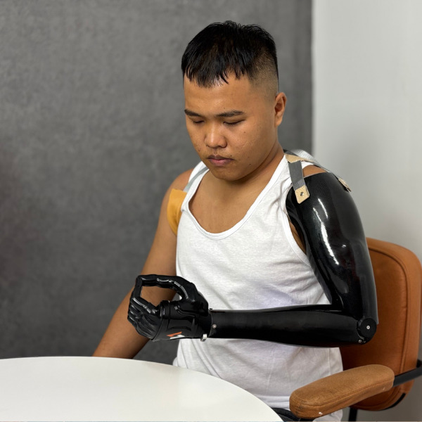
Weare_ Vulcan

The Importance of Personalizing EMG Signal in Prosthetics Control

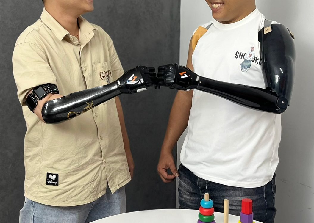
No two patients produce the same EMG signal. Yet many myoelectric fitting workflows still treat signal calibration as a checkbox rather than a clinical decision with predictable consequences for control reliability and device. Calibration is where the prosthetic control system learns what a specific patient’s muscle signals actually look like. Done well, it produces a device that responds predictably to deliberate commands and ignores incidental muscle activity. Done poorly or skipped in favour of default settings, it produces a device that feels unreliable, unpredictable, or simply too hard to use consistently. The gap between these two outcomes is not a function of the hardware or the myoelectric hand. It’s a function of how well the control system has been configured to the individual patient in front of the clinician. Why Individual Variation Makes One-Size Calibration Unreliable EMG signals are not standardised across patients. The amplitude, timing, and quality of the signals a person generates depend on a range of individual factors that vary significantly between patients, and within the same patient over time. Applying a fixed or population-average calibration to this variation produces control behaviour that fits no one particularly well. 01. Muscle mass and composition Patients with greater muscle mass or more superficial muscle bellies tend to generate higher-amplitude signals. Patients with atrophy, long-term limb loss, or complex tissue conditions may produce significantly lower amplitudes. A calibration designed for one produces the wrong sensitivity for the other. 02. Voluntary control experience New prosthetic users often produce inconsistent, variable signals as they learn to isolate and control residual muscle contractions. Experienced users may have well-established, reproducible patterns. Early calibration for a new user needs to accommodate inconsistency; later calibration can take advantage of the improved signal quality that develops with practice. 03. Residual limb condition Scar tissue, oedema, skin integrity issues, and limb volume fluctuation all affect signal quality and consistency. Two patients with the same amputation level and the same prosthetic hand may require very different calibration approaches because their residual limbs present entirely different signal environments. 04. Signal site location The specific location where a usable signal is found varies between patients and cannot be reliably predicted from anatomy alone. Moving even a small distance from the optimal site changes signal characteristics enough to require a different calibration response. 05. Fatigue and daily variation Signal amplitude in the same patient varies across the day as muscles fatigue, perspiration affects electrode contact, and limb volume changes. A calibration that works well in the morning fitting session may produce different control behaviour by the end of a working day. 06. Contralateral limb use In bilateral amputees or patients who rely heavily on compensatory upper body movement, crosstalk from muscles not intended to generate commands can introduce noise into the signal environment, requiring calibration settings that are more conservative to avoid false activations. These sources of variation don’t resolve on their own. They require active clinical attention during the calibration process, and they mean that calibration decisions made without considering the individual patient’s signal profile are likely to produce a suboptimal result. What Happens When Calibration Isn’t Personalised The consequences of generic or inadequate calibration appear in predictable patterns, most of which clinicians will recognise from patient feedback and return visit behaviour. False activations and unintended movement When calibration sets activation levels too low relative to a patient’s resting muscle activity, the hand responds to signals the patient didn’t intend as commands. This is one of the most commonly reported sources of frustration for myoelectric user. A device that moves when the patient doesn’t want it to, or that can’t be trusted in public settings where unexpected hand movement would be embarrassing. Non-response to deliberate commands When activation levels are set too high for a patient’s signal amplitude, which frequently happens with patients who have weaker signals due to atrophy or long-term limb loss, the device doesn’t respond to deliberate contractions. The patient has to over-effort to trigger a response, which is tiring, unreliable, and ultimately discouraging. Performance degradation across the day If calibration doesn’t account for signal variation due to fatigue, volume change, and perspiration, the control behaviour that feels acceptable in the morning may become noticeably less reliable by the afternoon. Patients who don’t understand why this happens often attribute it to the device being faulty, rather than recognising it as a calibration and signal quality issue. Excessive recalibration demand Patients who return repeatedly for calibration adjustments are often experiencing the downstream effects of initial calibration that wasn’t individualised sufficiently. Each visit addresses the symptom — imprecise control — without necessarily addressing the underlying cause, which is a calibration profile that doesn’t accurately reflect that patient’s signal characteristics. Device abandonment Unreliable control is a consistently cited factor in prosthetic device abandonment in the upper limb literature. Patients who cannot trust their device’s response, who experience it as unpredictable rather than responsive to their intent, are less likely to persist with use. Calibration that doesn’t fit the patient is a meaningful contributor to this outcome. Calibration as Patient-Specific Configuration Effective myoelectric calibration is patient-specific configuration, not a default setting applied uniformly, and not a one-time event at fitting that is assumed to hold indefinitely. It is a clinical process that benefits from the same systematic attention as other aspects of prosthetic fitting. Principles for More Effective Individual Calibration What This Means for Clinic Workflows Personalised calibration takes more time than applying a default. For clinics managing high patient volumes, that time pressure is real. However, the downstream cost of inadequate calibration, repeated return visits, frustrated patients, device abandonment, is consistently higher than the upfront investment in a more thorough initial calibration process. Clinics that treat calibration as a systematic clinical process, with defined steps, documented outputs, and scheduled review, tend to see fewer calibration-related return visits and more consistent patient-reported satisfaction with control reliability. The investment is at fitting; the return is in reduced follow-up burden and better long-term device use. How Vulcan approaches signal personalisation The Vulcan Myoband

The Importance of Personalizing EMG Signal in Prosthetics Control Read More »

Beyond Range of Motion: Why ROM Alone Isn’t Enough to Measure Functional Recovery

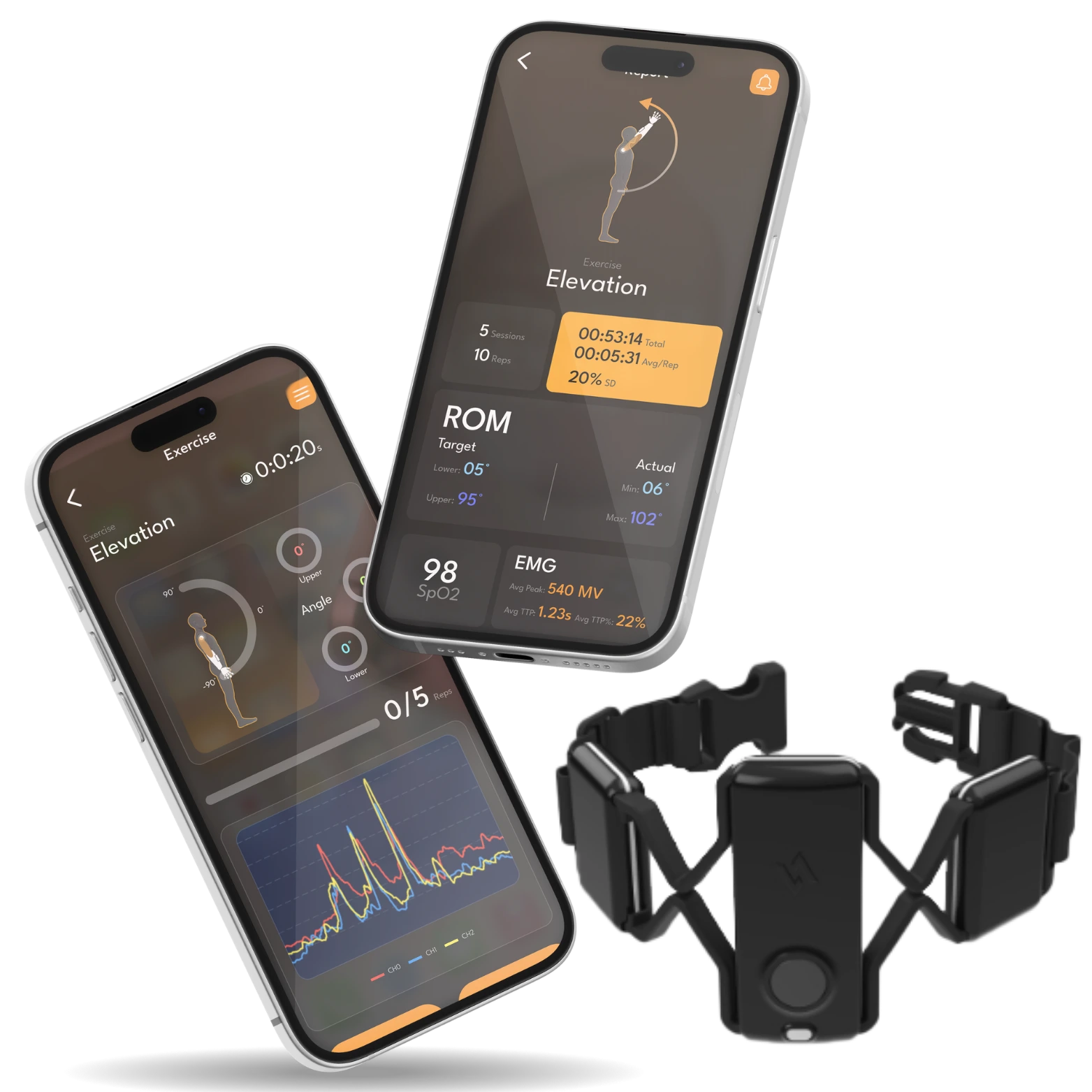
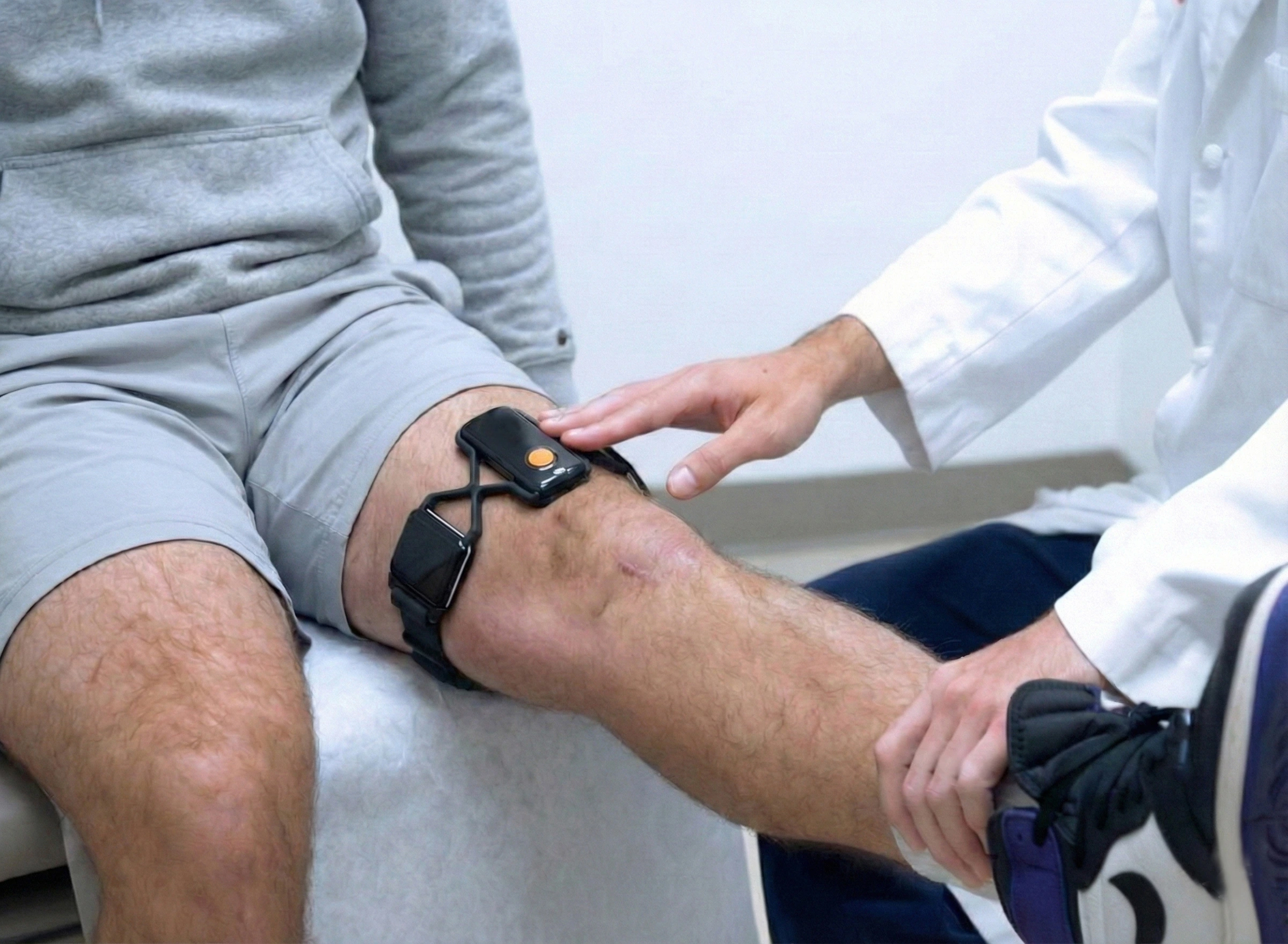
Range of motion is the most common metric in orthopedic rehabilitation. It’s also one of the most incomplete. Two patients can reach identical ROM targets and be at very different points in their recovery. A patient reaches 120 degrees of knee flexion at their six-week follow-up after total knee replacement. On paper, they’ve hit their target. Their surgeon marks the milestone. Their physical therapist moves the protocol forward. Then, three months later, the patient returns, still struggling with stairs, still avoiding certain movements, still not back to the activities they expected to resume. This isn’t an unusual story. And the disconnect at the center of it, between what range of motion tells clinicians and what’s actually happening in the patient’s recovery, is one of the more consequential limitations in how musculoskeletal outcomes are currently measured. What Range of Motion Actually Measures ROM assessment does one thing well: it tells you the angular range through which a joint can move. That’s a meaningful clinical data point. After surgery or injury, restricted joint movement is a clear indicator that something needs attention, and restoring motion is a legitimate early-stage rehabilitation goal. The problem isn’t that ROM is a bad measure. The problem is that ROM is frequently treated as a proxy for functional recovery — as though achieving a target angle is equivalent to recovering function. Research on orthopedic outcomes increasingly shows that these are not the same thing. Multiple studies across total knee replacement, ACL reconstruction, and rotator cuff rehabilitation have shown that muscle strength, neuromuscular activation, and movement quality are often stronger predictors of long-term function than ROM alone. (Mizner et al., Palmieri-Smith et al., Logerstedt et al.) What ROM tells you ✓ Whether the joint can move through a target angular range ✓ Whether there is gross restriction requiring intervention ✓ Whether a specific anatomical milestone has been reached ✓ Whether the patient can comply with a simple movement instruction What ROM doesn’t tell you The Muscle Activation Gap One of the most important limitations of ROM assessment is that it measures movement, but not the neuromuscular activity producing that movement. A patient may demonstrate adequate joint mobility while the muscles responsible for controlling that joint remain inhibited, weak, or poorly coordinated, a phenomenon well documented following orthopedic surgery. After total knee replacement, for example, patients often regain acceptable knee flexion before quadriceps activation fully recovers. Although ROM targets may be achieved, persistent quadriceps deficits can remain for months and are associated with poorer stair negotiation, reduced walking performance, and lower patient-reported function. This illustrates a broader principle: joint mobility and functional recovery do not always progress at the same rate. Movement Quality: What Angle Measurements Miss Beyond muscle activation, ROM measurements are blind to how a movement is being performed. Two patients can achieve the same joint angle through very different movement strategies, one recruiting the correct muscle groups in a coordinated pattern, one compensating through adjacent structures in a way that distributes load inefficiently and may create new problems over time. Compensation patterns are particularly common in post-surgical patients. They often develop gradually and quietly, the patient finds a way to accomplish the assessed movement that satisfies the clinical benchmark while avoiding the tissue or muscle group that is not yet ready to bear full load. The ROM number looks fine. The movement quality does not. Four Dimensions ROM Doesn’t Capture Functional recovery is multidimensional. ROM captures one of those dimensions. Clinicians making recovery and clearance decisions ideally want to understand all of them, and the gap between what ROM provides and what comprehensive recovery assessment would provide is substantial. The core problem Range of motion is a necessary but insufficient indicator of recovery. It answers one question — can the joint move through this range? — while leaving the questions that most directly predict functional outcomes unanswered. When ROM is used as a primary recovery milestone rather than one data point among several, it creates a false ceiling: patients who reach target ROM are classified as recovered when recovery may still be substantially incomplete. What Comprehensive Recovery Assessment Looks Like The clinical shift being discussed across orthopedic and rehabilitation medicine is not away from ROM, it’s toward using ROM alongside objective data on muscle activation, movement quality, and physiological markers that together provide a more complete picture. For clinicians, this has practical implications at several stages of rehabilitation: Early recovery — weeks 1 to 6 ROM is most useful here. Gross restriction is the primary concern, and ROM targets provide clear early benchmarks. However, monitoring muscle activation alongside ROM from the start identifies patients whose neuromuscular recovery is lagging behind their joint mobility, a pattern that predicts later functional deficits if not addressed early. Mid-recovery — weeks 6 to 12 This is where ROM alone becomes most misleading. Patients frequently reach ROM targets while muscle activation and movement quality remain significantly impaired. Protocols advanced based on ROM milestones at this stage can miss the patients who are hitting the numbers but not the function. Return-to-activity clearance Return-to-sport, return-to-work, and return-to-daily-activity decisions made on ROM data alone have the highest risk of misclassification. Research across multiple surgical categories has linked clearance decisions based purely on ROM or time-based criteria to elevated re-injury rates and persistent functional limitations. How Vulcan Approaches This This is the clinical problem the Vulcan MSK Sensor System is designed to address. Rather than replacing ROM assessment, the system is intended to sit alongside it — capturing muscle activity, motion quality, and physiological signals during in-clinic assessment and home rehabilitation to give clinicians a more complete picture of where recovery actually stands. Currently in clinical pilotsThe system supports both in-clinic biomechanical assessment — where a clinician places the wearable sensor and captures objective data during prescribed movements — and remote home monitoring, where the patient performs their rehabilitation protocol at home and clinicians access objective adherence and recovery data through the platform. Active investigator-initiated studies at orthopedic centers are currently evaluating the system’s clinical utility in Total

Beyond Range of Motion: Why ROM Alone Isn’t Enough to Measure Functional Recovery Read More »

Why Myoelectric Hand Users Often Can’t Wear Soft Liners and What’s Changing

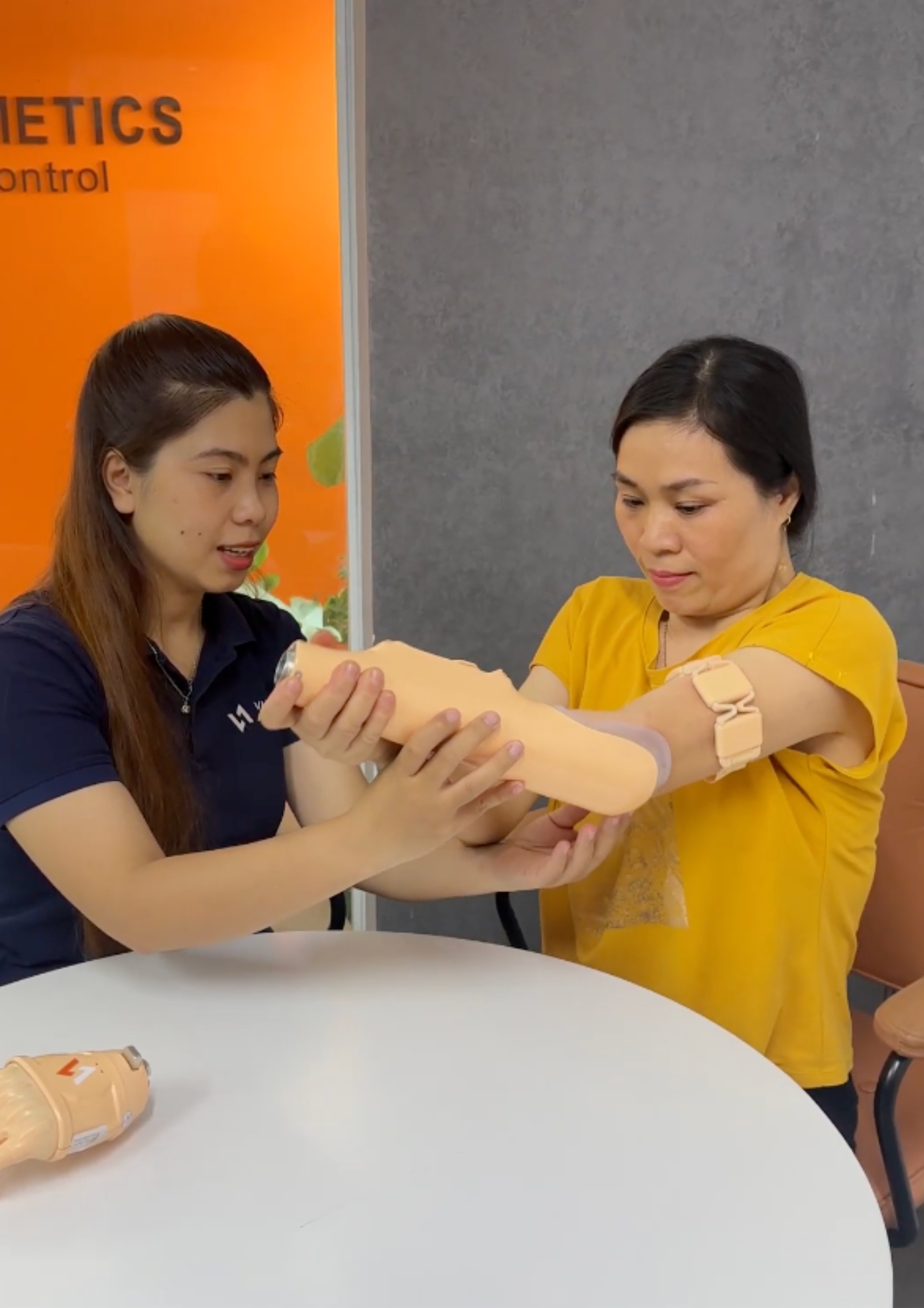
Soft liners improve comfort, suspension, and skin protection for prosthetic users. For most myoelectric hand users, they’ve historically come with a catch: the electrodes need direct skin contact that a liner prevents. Soft liners have been used in lower limb prosthetics for decades. In upper limb prosthetics, adoption has grown  but for users of myoelectric hands, a long-standing obstacle has prevented many patients from accessing the same comfort benefits: the liner sits between the skin and the electrodes that the control system depends on. This isn’t a minor inconvenience. It’s a clinical trade-off that forces many patients and clinicians to choose between comfort and reliable control and until recently, the options for resolving that trade-off were limited and technically demanding. Why Liners Matter for Upper Limb Users The interface between the residual limb and the socket is one of the most consequential factors in long-term prosthetic use. Even a well-designed prosthetic hand becomes difficult to use consistently if the socket causes discomfort, skin breakdown, or inconsistent fit across the course of a day. Soft liners, typically made from silicone, urethane, or thermoplastic elastomeric materials, are reported to offer several clinical benefits that address common socket fit challenges.1 Despite these benefits, upper limb myoelectric users cannot wear a liner because the EMG electrode must touch the skin to capture the myoelectric signal. Therefore, upper limb users miss out on the comfort and suspension benefits of the liner, while also not being able to use prosthetic socks for limb volume management.3 The Core Problem: Electrodes Need Skin Contact The clinical consequence is that many myoelectric users who would benefit from a liner, patients with skin integrity concerns, volume fluctuation, or discomfort with hard socket interfaces, have had to go without one. And the workarounds that do exist have typically involved compromises of their own. How the Field Has Tried to Solve It Several approaches have been developed over the years to allow liner use with myoelectric systems. Each has moved in a useful direction, but each also carries trade-offs that have limited broader clinical adoption. A Different Starting Point: Sensing Outside the Socket A different approach is to remove the electrodes from the socket entirely. When signal acquisition happens outside the socket, through a wearable sensor band worn on the residual limb, transmitting wirelessly to the prosthetic hand, the liner-electrode conflict doesn’t arise, because there are no socket-embedded electrodes to conflict with. It is worth noting that this approach involves trade-offs of its own. A wearable external band adds a donning step and requires consistent positioning on the limb. The clinical suitability of any approach, socket-embedded, liner-integrated, or wearable external, will depend on the individual patient’s anatomy, activity level, and clinical context. What changes with an external wearable is that the liner question becomes independent of the electrode question, giving clinicians more flexibility to address both on their own terms. What This Means Clinically For CPOs working with patients who have skin integrity concerns, volume fluctuation, or significant discomfort with hard socket interfaces, the liner question is a real and recurring clinical consideration. The field has been working on it for years, through liner modifications, embedded electrode systems, and now wireless architectures, and the solutions are becoming more clinically practical. The underlying question remains worth keeping in mind: if a patient would benefit from a liner, what would it take to give them one without compromising their myoelectric control? The answer increasingly depends less on modifying the liner to accommodate the electrode, and more on rethinking where the electrode needs to be in the first place. Learn more about how the Vulcan Myoband’s wearable, socket-independent design supports a wider range of fitting configurations. Reference 1. Reissman T, Halsne E, Lipschutz R, Miller L, Kuiken T. “A novel gel liner system with embedded electrodes for use with upper limb myoelectric prostheses.” PLOS One, June 2018. DOI: 10.1371/journal.pone.0198934 — Benefits of liners (skin protection, cushioning, suspension, adjustability) are cited from general liner literature within this paper’s introduction, not primary findings of this study. 2. Reissman et al., 2018 (same as ref 1). Direct finding from 8-subject home trial: “Subjects preferred the liner prototype (p = 0.008) over their own system in the clinical areas of comfort, suspension, function, and, especially, ease of use.” 3. ASTERISK Study Protocol. ClinicalTrials.gov NCT06821412. “Wireless Prosthetic Control Effectiveness Study.” Accessed June 2026. Source of quote: “upper limb prosthesis users with myoelectric control cannot wear a liner as the electromyogram (EMG) electrode must touch the skin to transduce the myoelectric signal.” 4. Cited approaches (piercing liner with electrode domes; cutting holes in liner) documented in Reissman et al. 2018 introduction, referencing earlier clinical literature on liner modification approaches.

Why Myoelectric Hand Users Often Can’t Wear Soft Liners and What’s Changing Read More »

EMG Signal Visualization in Myoelectric Fitting: A Clinical Decision-Making Guide

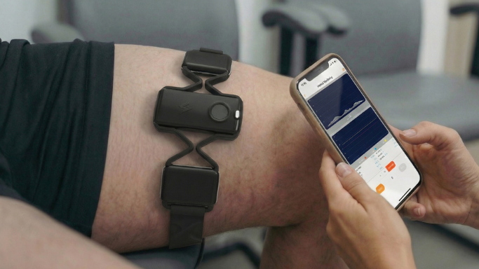
When EMG signal data becomes visible in real time, fitting stops being a trial-and-error process. This is what clinicians can see, what it means, and when it changes the decision they make next. Myoelectric fitting has traditionally been an experience-dependent process. Signal quality is assessed through palpation and patient feedback, electrode placement is adjusted by feel, and calibration is confirmed when the hand moves the way the clinician expects. There’s expertise in that approach, but there’s also a ceiling on what it can reliably tell you. When EMG signal data is visible in real time, waveforms on screen, contraction timing captured as it happens, motion data updating continuously, the clinical picture changes. Decisions that were previously made by inference can be made on observation. This article is about what that visibility actually shows, how to read it usefully, and where it changes the decisions that matter in fitting and follow-up. What Fitting Looks Like Without Signal Visibility In a conventional myoelectric fitting, a clinician’s understanding of the patient’s EMG signal is largely indirect. They can feel for muscle activity through palpation. They can watch the hand respond during calibration and draw conclusions from the response. They can ask the patient to contract and observe whether the movement is what they expected. This works reasonably well when the patient has clear, strong signals and cooperative anatomy. It becomes progressively harder to rely on when signals are weak, inconsistent, or distributed across the limb in ways that aren’t easy to locate by palpation alone. Without real-time signal visualization With real-time signal visualization ✓ Signal quality confirmed at each candidate site before placement ✓ Contraction strength and timing visible directly ✓ Calibration based on measurable activation data ✓ Signal trends reviewable at follow-up ✓ Candidacy confirmed or ruled out objectively before fabrication Five Clinical Decisions That Signal Visualization Directly Supports Real-time EMG visibility isn’t a single-use tool. It supports different clinical questions at different stages of the fitting process — from the very first evaluation through to long-term follow-up. 01. Pre-fabrication candidacy assessment Before investing time in socket fabrication, visualizing actual EMG signal output allows clinicians to confirm whether a patient can generate consistent, usable signals and from which sites. Patients who appear marginal on palpation alone may produce viable signals when assessed with objective data.Reduces misclassification risk. Reduces misclassification risk 02. Signal site selection Comparing signal quality across multiple candidate sites — amplitude, consistency, noise ratio — allows for more systematic site selection rather than relying on the first viable location found. Particularly useful for patients with atrophy, scar tissue, or short residual limbs where signal distribution may be less predictable. Reduces myosite hunting time 03. Calibration quality confirmation Visual feedback during calibration lets the clinician see whether contraction and relaxation levels are being captured cleanly, and whether the patient’s activation pattern is consistent enough to support reliable control. Calibration that looks correct on the device may still have underlying signal variability worth addressing. Evidence-based calibration 04. Patient engagement in training When patients can see their own muscle activity in real time, motor relearning accelerates. Visual biofeedback, the patient seeing their contraction register on screen, supports neuromuscular re-engagement in ways that verbal cues alone typically don’t. This is particularly relevant for long-term amputees returning to myoelectric use. 05. Follow-up signal trending Reviewing stored signal data at follow-up appointments gives clinicians a longitudinal view of how a patient’s activation patterns have changed. Signals that were marginal at fitting may have strengthened with use and training, or may have degraded, pointing to limb changes that need to be addressed. A Practical Signal Assessment Workflow The following steps reflect general clinical practice for EMG signal assessment during myoelectric fitting, applicable regardless of which visualization tool or sensor system a clinic uses. The underlying principle is consistent: establish a baseline, compare sites systematically, test under real conditions, and document for follow-up. 1. Establish a resting baseline first Before asking the patient to contract, observe the signal at rest. The resting baseline tells you the noise environment, and how clean the voluntary contraction signal will need to be to be distinguishable from it. 2. Map multiple candidate sites systematically Rather than confirming the first viable signal and moving on, compare at least two to three candidate sites before selecting. The best available site may not be where you’d expect it from anatomy alone — particularly in patients with long-term limb loss or tissue changes. 3. Test under movement, not just at rest Have the patient perform arm elevation, reaching, and load-bearing movements while monitoring signal quality. A site that looks clean in the static assessment may show significant positional sensitivity or crosstalk under dynamic conditions — which will matter the moment the patient uses the device at home. 4. Use the signal view during calibration, not just before it Keeping the signal display active during calibration lets you see whether the patient’s activation pattern is settling into consistency or still varying. Inconsistency during calibration is easier to address before the settings are confirmed than after the patient has gone home. 5. Document the signal profile for follow-up comparison A signal profile captured at fitting becomes a clinical reference point. At follow-up, comparing current signal data against the initial session tells you whether activation is strengthening, degrading, or stable, information that shapes the next set of clinical decisions. Note: the specific steps and tools vary significantly between control systems. The workflow below applies primarily to threshold-based myoelectric systems using real-time signal visualization, the approach differs for pattern recognition systems, which have their own calibration and training protocols. Signal Visualization in the Vulcan App The Vulcan Myoband is designed to stream EMG data to the Vulcan app during fitting and calibration, giving clinicians a live view of muscle signal amplitude, contraction timing, and motion data during every stage of the fitting and calibration process. The signal history can be reviewed at follow-up appointments, supporting the kind of longitudinal tracking described in this article.

EMG Signal Visualization in Myoelectric Fitting: A Clinical Decision-Making Guide Read More »

How Poor Adherence to Home Exercise Programs Derails MSK Recovery

Prescribing the right exercises is only half the job. What happens when the patient leaves the clinic and whether they actually do them, is where a meaningful share of orthopedic rehab outcomes are quietly won or lost. Home exercise programs are one of the most widely used tools in orthopedic rehabilitation and one of the least reliably followed. The exercises themselves are rarely the problem. Whether they actually get done, consistently, over the weeks between appointments, is where recovery often quietly comes off track. This isn’t a minor compliance footnote. Research on home exercise program (HEP) adherence puts non-adherence as high as 70% in some musculoskeletal populations¹, meaning that for a meaningful share of patients, the prescribed dose of exercise is never actually delivered, regardless of how well the program was designed. ¹ Based on published research on HEP adherence in musculoskeletal populations. How Big the Gap Actually Is 35% of physical therapy patients reported to fully adhere to their prescribed home exercise program Source: Industry analysis of PT adherence patterns (SPRY, 2025) — directional estimate, not peer-reviewed primary data 70% non-adherence reported in some musculoskeletal populations, including patients with neck pain Source: Himler et al., Musculoskelet Sci Pract, 2023 A 2018 study published in the Journal of Orthopaedic & Sports Physical Therapy used concealed accelerometers to compare what patients said they did against what they actually did. The gap between self-reported and objectively measured adherence was consistent enough to suggest that clinics relying purely on patient recall may be working with an incomplete picture, and that the actual dose of exercise being delivered may differ meaningfully from what the chart reflects.⁴ Why Patients Don’t Follow Through The research on HEP non-adherence is fairly consistent on what the actual barriers tend to be, and “laziness” or simple forgetfulness rarely top the list. Notably, depression has strong supporting evidence as a barrier to adherence⁵, and pain affecting more than one body region has been linked specifically to motivational, rather than physical, barriers to completing a HEP.³ Adherence isn’t a single, uniform behavior; it’s shaped by a mix of physical, psychological, and logistical factors that differ from patient to patient. Why This Matters for Outcomes, Not Just Compliance Adherence to a HEP directly affects the overall dose of therapeutic exercise a patient receives. Poor adherence can mean a patient is effectively under-dosed relative to what their recovery actually requires³ , and research has associated HEP adherence with superior functional outcomes.³ In other words, this isn’t just a workflow or billing concern. It’s a meaningful lever on whether a patient recovers on the timeline their treatment plan assumes. What Actually Moves the Needle The research points less toward stricter enforcement and more toward addressing the specific barrier in front of a given patient. The Underlying Issue Most of these barriers — pain, uncertainty, low confidence, and an overwhelming program — are addressable. The challenge is that they’re rarely visible to the clinical team until adherence has already broken down and outcomes have started to lag. The gap isn’t a lack of good HEP design. It’s a lack of visibility into what’s actually happening in the weeks between appointments. This is one part of a broader pattern this series has explored: a significant share of musculoskeletal recovery happens where clinicians can’t directly observe it. Vulcan has been studying this problem closely through the development of the Vulcan MSK Sensor System, a wearable sensor and analytics platform currently being piloted to give clinicians more continuous visibility into recovery — including the adherence patterns that are otherwise invisible between visits. Learn how the wearable sensor and analytics platform are being piloted across hospital, home-care, and clinical practice settings. References 1. Himler P, Lee GT, Rhon DI, Young JL, Cook CE, Rentmeester C. “Understanding barriers to adherence to home exercise programs in patients with musculoskeletal neck pain.” Musculoskeletal Science and Practice, 2023. 2. “Physical Therapists’ Assessment of Patient Self-Efficacy for Home Exercise Programs.” International Journal of Sports Physical Therapy, 2025. 3. “Adherence and Barriers to Home Exercise Program Participation in Adults With Musculoskeletal Pain.” Archives of Physical Medicine and Rehabilitation / ScienceDirect, 2022. 4. “Self-reported Home Exercise Adherence: A Validity and Reliability Study Using Concealed Accelerometers.” Journal of Orthopaedic & Sports Physical Therapy, 2018. 5. “Adherence to Home Exercise Programs.” Physiopedia, summarizing Bassett SF, “Barriers to treatment adherence in physiotherapy outpatient clinics: A systematic review,” Manual Therapy, 2010. 6. “Boost Exercise Adherence” — industry analysis citing PT adherence research, SPRY, 2025. (35% full-adherence figure; treated as directional, not peer-reviewed primary data.)

How Poor Adherence to Home Exercise Programs Derails MSK Recovery Read More »

The Post-Discharge Blind Spot

Most musculoskeletal recovery happens outside the clinic. As Remote Therapeutic Monitoring gains traction under 2026 reimbursement changes, the question clinicians are asking is the same one they’ve always asked: what’s actually happening to my patient between appointments? A patient leaves the hospital after a knee replacement. The surgery went well. They’re sent home with a printed exercise sheet, a follow-up appointment in six weeks, and their surgeon’s best hope that they’ll do as instructed. What happens between now and that follow-up is, for the most part, invisible. The surgeon won’t know if the patient completed their exercises. They won’t know if pain on day twelve caused the patient to quietly stop moving. They won’t see a compensatory gait pattern forming over three weeks of guesswork. By the time the follow-up appointment arrives, the recovery trajectory may already have drifted and correcting course is far harder than staying on it from the start. This is the post-discharge blind spot. It isn’t a failure of clinical skill. It’s a structural gap in how musculoskeletal recovery is currently tracked and in 2026, it’s becoming one of the most actively discussed problems in orthopedic and rehabilitation care. The Recovery Clinicians Don’t See Surgical technique, implant design, and evidence-based rehab protocols have all advanced considerably over the past two decades. One thing hasn’t changed much: the visibility window clinicians have into how a patient is actually doing. Recovery from major musculoskeletal procedures is rarely a brief event. Total knee arthroplasty involves meaningful recovery over many months. Rotator cuff repair can extend well beyond that. ACL reconstruction requires sustained rehabilitation over a year or more. During that stretch, patients typically see a physical therapist a few times a week at most  and receive no direct clinical observation for the rest of it. That gap accounts for the large majority of the total recovery period. It’s also where the most important decisions about whether a patient is on track, or quietly falling behind, would ideally be made. Five Questions Clinicians Still Can’t Reliably Answer The information gap created by the post-discharge blind spot shows up as a set of clinical questions that remain stubbornly difficult to answer with confidence, not edge cases, but central to how rehabilitation decisions get made. Why Self-Reporting Doesn’t Close the Gap The default tool for understanding what happens between visits is the patient themselves outcome questionnaires, pain scales, and a conversation at the follow-up appointment. These are useful, but they were never designed to capture six weeks of daily variation. Research on patient recall:  most of it from pain assessment literature, points to a real but modest effect: people’s memory of how they felt over a period tends to be disproportionately shaped by the most intense moments and the most recent ones, rather than an even average of the whole stretch. The effect isn’t large enough to call self-reporting unreliable, but it’s a reminder that what a patient describes at a follow-up visit is a reconstruction, filtered through memory, not a continuous record of what actually happened. Self-reporting also can’t capture what a patient doesn’t notice in the first place. Subtle changes in movement quality, early signs of muscle compensation, or a gradual decline in activation are often invisible to the patient, especially early in recovery when proprioception itself may be compromised. These are also frequently the signals that matter most for catching a problem early. Why This Conversation Is Happening Now Remote Therapeutic Monitoring has existed as a Medicare billing category since 2022, but 2026 marks a meaningful shift. In its CY 2026 Physician Fee Schedule Final Rule, CMS added two new CPT codes for musculoskeletal RTM, 98985 and 98979, specifically designed to lower the eligibility threshold. Previously, qualifying for RTM reimbursement required at least 16 days of data collection and 20 minutes of provider management time per month; the new codes recognize shorter monitoring periods (as few as 2–15 days) and shorter management windows (10–19 minutes), making RTM workable for a wider range of patients and practice types. What We’re Seeing in Early Pilots This is a problem Vulcan has been studying closely through the development of the Vulcan MSK Sensor System, a wearable sensor and companion analytics platform designed to give clinicians objective visibility into recovery, including the periods between scheduled visits. The system combines a wearable sensor band with a clinician-facing analytics platform, capturing muscle activity, motion, and effort to give a more continuous picture of recovery than periodic in-clinic assessment alone allows. The post-discharge blind spot isn’t an unsolvable feature of musculoskeletal care, it’s an information problem. And it’s one the industry, helped along by changing reimbursement incentives, is finally starting to take seriously. Learn how the wearable sensor and analytics platform are being piloted across hospital, home-care, and clinical practice settings.

The Post-Discharge Blind Spot Read More »

EMG Signal Acquisition in Myoelectric Prosthetic Fitting: Clinical Challenges and Practical Solutions for CPOs

Before a patient can successfully control a myoelectric prosthesis, clinicians must first solve one of the most critical and often unpredictable aspects of the fitting process: acquiring reliable EMG signals. While prosthetic technology continues to evolve, the quality of myoelectric control still depends heavily on what happens during signal acquisition. The effectiveness of control training, calibration, and long-term device performance all begin with the ability to consistently identify and capture usable muscle activity. Why Is EMG Signal Acquisition Challenging? In theory, acquiring a usable EMG signal is straightforward: place electrodes over the target muscle, ask the patient to contract, observe the output. In practice, the process is confounded by a range of patient-specific, physiological, and anatomical variables that make no two fittings alike. 1. Residual Limb Variability Limb length, tissue composition, skin condition, and scar tissue distribution differ significantly between patients and within the same patient over time as the limb matures post-amputation. 2. Muscle Atrophy and Reduced Signal Strength Patients with long-term limb loss often present with significantly reduced muscle mass in the residual limb. Atrophied muscle produces weaker signals with lower signal-to-noise ratios, making reliable acquisition technically demanding. 3. Scar Tissue Interference Post-surgical scar tissue increases impedance at the skin surface, attenuating signal amplitude and introducing inconsistency in electrode contact quality, particularly problematic for traditional socket-embedded electrodes. 4. Signal Crosstalk In short residual limbs or limbs with limited tissue differentiation, electrical activity from adjacent muscles can contaminate the target signal, producing false activations and unpredictable control responses. 5. Variability in Voluntary Control New prosthetic users often lack consistent voluntary muscle activation patterns. Signals vary in amplitude, timing, and repeatability between sessions, particularly in the early weeks post-fitting. 6. Dynamic Changes During Daily Use Traditional socket-embedded electrodes are positioned for a specific limb configuration. Limb volume changes from perspiration, weight fluctuation, or tissue adaptation can shift electrode-skin contact and degrade signal quality between fittings. These variables do not present in isolation. A typical complex patient may exhibit weak baseline signals, compromised tissue quality, and limited voluntary control simultaneously, requiring the CPO to manage all three in a single fitting session. How Poor EMG Signal Acquisition Affects Clinical Outcomes Signal acquisition problems are not always obvious during fitting appointments. Patients may demonstrate acceptable control while seated in a controlled clinical environment yet struggle significantly once they return to the unpredictable demands of daily life. Poor signal quality can contribute to: Every unsuccessful fitting carries hidden costs, not only in clinician time and resources but also in the patient’s trust in the technology itself. A Systematic Approach to EMG Signal Assessment Managing signal acquisition complexity begins before the electrode is placed. A structured pre-fitting assessment protocol reduces the likelihood of downstream control problems and gives the CPO a reproducible framework for evaluating each patient’s signal landscape. 1. Residual Limb Mapping Before electrode placement, systematically assess the residual limb for tissue quality, scar tissue location, muscle belly palpation, and approximate signal site candidates. Document findings for reference across sessions. This step is frequently abbreviated under time pressure — at significant downstream cost. 2. Baseline Signal Characterization Use real-time EMG visualization to establish baseline signal amplitude, resting noise levels, and voluntary contraction quality across candidate sites before committing to electrode placement. Compare multiple sites systematically rather than relying on the first adequate signal identified. 3. Dynamic Signal Testing Assess signal quality under movement conditions, not just at rest. Have the patient perform arm movements, elevation changes, and load-bearing tasks while monitoring signal consistency. A signal that is stable in static testing often degrades significantly under dynamic conditions. 4. Patient-Specific Threshold Calibration Calibrate activation thresholds to the individual patient’s signal profile — not to a population average. Patients with weaker signals require lower thresholds and higher gain settings; patients with strong signals or crosstalk may need more restrictive thresholds to prevent false activations. 5. Session-to-Session Reassessment At the second fitting session, re-evaluate signal quality with the same protocol used in session one. Significant variation between sessions is diagnostically important, it identifies patients whose signal profile is not yet stable and who will require ongoing monitoring and recalibration. How Multi-Sensor Coverage Changes the Acquisition Equation The choice of control approach depends on the patient’s functional goals and signal profile. But regardless of which approach a clinician selects, the quality of EMG signal acquisition upstream determines how well any system will perform. Advanced control technologies depend on consistent and repeatable signal input to function as intended. When the underlying signal is weak, inconsistent, or contaminated by crosstalk, no control layer can fully compensate. Traditional myoelectric systems rely on precise placement of one or two electrodes over specific muscle bellies. The clinician must palpate the residual limb while the patient performs repeated contractions to locate a single viable motor point: a process clinicians often describe as “myosite hunting.” In ideal anatomy, this works well. In residual limbs with scar tissue, atrophy, or limited muscle differentiation, it can consume a disproportionate share of fitting time before device viability is even confirmed. The Vulcan control system calibrates automatically to each patient’s individual muscle activity through the Vulcan app in roughly 60 seconds. Commands are transmitted wirelessly to the prosthetic hand, with no embedded wiring inside the socket to maintain. What Consistent Signal Acquisition Makes Possible The clinical benefits of reliable EMG signal acquisition extend well beyond fitting day. When signal quality is stable and reproducible, everything downstream becomes more manageable: control training is faster, patient confidence builds earlier, recalibration frequency drops, and long-term device adoption improves. The goal of signal acquisition is not to obtain the strongest signal. It is to obtain the most reliable signal for that patient, one that will perform consistently across the full range of conditions their daily life presents. That requires a systematic approach, the right assessment tools, and a sensing architecture designed for real-world variability rather than clinical ideal conditions. See How Vulcan Approaches Signal Acquisition Explore how the Myoband’s multi-sensor architecture simplifies clinical fitting across complex patient profiles.

EMG Signal Acquisition in Myoelectric Prosthetic Fitting: Clinical Challenges and Practical Solutions for CPOs Read More »

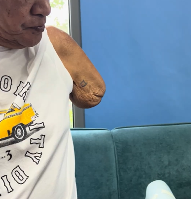
A close-up side profile of an elderly individual with an upper-limb amputation above the elbow. The residual limb shows visible skin wrinkling and signs of muscle atrophy. A small, simple square tattoo is visible on the side of the limb.

When Muscles Change: How Vulcan Supports Patients With Muscle Atrophy and Long-Term Limb Loss

One of the less-discussed challenges in upperlimb prosthetic care is what happens to muscle tissue over time and what that means for myoelectric control. For many patients, the residual limb doesn’t stay the same after amputation. Muscles that aren’t regularly activated begin to atrophy. Fatty and fibrous tissue accumulates. The signals that a prosthetic system needs to read become quieter, less stable, and harder to interpret. In some long-term cases or congenital presentations, those signals may be extremely faint from the outset. Traditional electrode systems weren’t designed with this in mind and for a significant portion of the prosthetic population, that’s a real limitation. What Happens to EMG Signals as Muscles Atrophy Following amputation, the residual limb goes through a prolonged process of biological change. The most significant shifts typically occur in the first 6 to 18 months, but remodeling can continue indefinitely particularly in patients who don’t use a prosthesis early or who have limited physical activity. These changes affect EMG signal quality in several ways: Signals become less stable Atrophied muscle has fewer active motor units firing in a coordinated way. The result is a signal that fluctuates, making it difficult for patients to maintain a consistent contraction and for the system to interpret intent accurately. Tissue changes increase impedance As fatty and fibrous tissue accumulates between the muscle and the electrode, signal conduction attenuates. Electrode contact becomes less consistent, particularly as limb shape continues to change. Noise becomes a bigger problem When signal amplitude is low, the ratio of useful signal to background noise deteriorates. Crosstalk from adjacent muscle groups increases, and the risk of the system misreading a signal or missing one entirely goes up. Why Conventional Electrode Systems Fall Short Traditional dual-electrode setups depend on two things: accurate placement over a defined motor point, and signals strong enough to reliably cross a fixed threshold. In a healthy, recently-fitted patient, that’s a reasonable assumption. In a patient with significant atrophy, neither condition may hold. Motor points may no longer be clearly defined. Signals may never consistently reach the threshold needed to trigger a command. The system that worked at fitting may become unreliable months later as the limb continues to change. For long-term amputees, non-users returning to prosthetic care, or individuals with congenital limb differences, this creates a meaningful barrier to myoelectric control — not because the patient can’t generate signals, but because the system can’t detect and interpret the ones they have. How Vulcan Approaches It Differently Rather than requiring a strong signal from a fixed location, the Vulcan Myoband is designed to work with what’s available across the full residual limb. The Myoband encircles the limb with multiple sensors, so it isn’t dependent on a single site holding up over time. If muscle activity in one area is reduced, the system continues to draw on signal from elsewhere around the limb, meaning a single atrophied or fibrotic region doesn’t determine whether control is possible. Reading Signal, Not Just Strength Conventional systems work on a simple rule: if the signal crosses a set strength, trigger a command. For patients with weak or atrophied muscle signals, that level may never be consistently reachable. The Vulcan Myoband is built to recognize meaningful muscle activity even when it’s faint, calibrating to what each patient’s signal actually looks like, rather than expecting every patient to produce the same signal strength. Built for Faint and Inconsistent Signals Patients with significant atrophy often generate muscle activity too subtle for conventional electrodes to register. The Myoband is designed to pick up on activity at this level, even when individual signals are small or inconsistent. This gives clinicians a clearer picture of where viable muscle activity exists across the residual limb, useful information when deciding which sites to focus on during training, especially in complex or long-term cases. A Three-Step Clinical Protocol for Atrophy Cases For patients with muscle weakness or long-term limb loss, Vulcan’s approach follows a structured sequence: 1. Calibration The Vulcan app learns the patient’s current contraction levels during initial setup, even if those contractions are extremely faint. Rather than requiring the patient to meet a fixed standard, the system is calibrated individually, starting from where the patient’s signal actually is. 2. Visual feedback The patient can see their muscle activity in on screen. This isn’t just reassuring, it’s clinically meaningful. Visual feedback helps the brain reconnect with and learn to control muscles that may have had limited use for months or years, supporting the neuromuscular re-engagement that underpins effective prosthetic training. 3. Physical conditioning Alongside device training, daily massage and isometric exercises help maintain remaining muscle fiber density and improve local blood flow. Preserving what muscle tissue remains makes a measurable difference to long-term signal quality and control consistency. Why This Matters for Clinical Practice Muscle atrophy is not a niche presentation. It affects long-term amputees, late fitters, patients who’ve had previous prosthetic abandonment, and individuals with congenital limb differences. It’s also a progressive condition, meaning patients who are well-controlled at fitting may become harder to manage over time if the system can’t adapt. A control approach that reads signal patterns rather than raw amplitude, captures data across the full limb rather than fixed points, and adapts its baseline to the individual patient is better positioned to serve this population both at the point of fitting and across the years that follow.

When Muscles Change: How Vulcan Supports Patients With Muscle Atrophy and Long-Term Limb Loss Read More »

Adaptive Control: How Vulcan Keeps Prosthetic Response Consistent in Daily Life

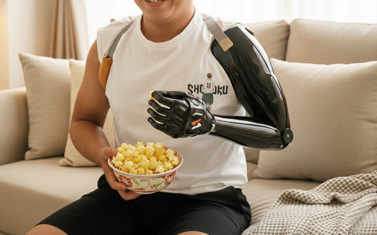
Adaptive control keeps the logic clinicians and patients already know: a lighter contraction opens the hand, a stronger one closes it. It’s intuitive, easy to calibrate, and easy to learn, which is exactly why it remains the most widely used control method in myoelectric prosthetics. What the Vulcan Myoband changes is how that logic holds up once the patient leaves the clinic and moves through a normal day. Why EMG Signals Don’t Stay Consistent The difficulty isn’t the control method itself. It’s that EMG signals naturally vary with fatigue, posture, and electrode contact and a fixed calibration has no way to account for that variation. A calibration that feels stable in the clinic can behave differently at home, not because anything was set up incorrectly, but because the signal environment has changed. For clinicians, this often means repeated adjustments at follow up. For patients, it can mean a device that feels less predictable than expected, which over time affects confidence and daily use. How Vulcan Handles It Differently Vulcan keeps the same basic control logic: lower activation for one action, higher for another because it works and patients understand it. What’s different is how the system holds up once real-world conditions come into play. The Myoband doesn’t just read muscle signals in isolation. It also accounts for what the arm itself is doing so the system can tell the difference between a deliberate command and ordinary movement. Reaching for an object naturally tightens stabilizing muscles something a fixed system can mistake for a command. The Myoband is built to tell the difference, so the hand responds to deliberate movement, not incidental muscle activity. Same control logic patients are already familiar with, one that adapts to real conditions rather than assuming they stay constant. What the Data Tells Clinicians Over Time Because the system tracks performance over time, clinicians build a picture of how each patient’s muscle activity evolves. Are activation levels becoming more consistent? Is the patient needing to contract harder than before? That kind of longitudinal insight is difficult to get from a conventional setup, where calibration changes happen by feel and little gets recorded systematically. Having it available makes follow-up conversations more grounded and gives rehabilitation teams something concrete to work from when planning training adjustments. What It Means in Practice For patients: Day-to-day control feels more predictable. Fewer unexpected hand movements, less frustration when the device doesn’t respond as expected. The learning curve stays manageable because the underlying logic doesn’t change, it just holds up better under real conditions. For clinicians and CPOs: The calibration process stays familiar. The difference is fewer return visits for calibration adjustments, and more confidence that settings will hold between appointments. For rehabilitation teams Longitudinal signal data adds a layer of objectivity to recovery tracking that conventional systems don’t easily provide.

Adaptive Control: How Vulcan Keeps Prosthetic Response Consistent in Daily Life Read More »

A clinician and a patient with a transradial amputation evaluating real-time EMG signals. The patient wears a Vulcan wireless myoband on the residual limb, while a smartphone on a stand displays a visual graph of muscle activation thresholds.

Real-Time EMG and IMU Visualization in Prosthetic Care: How the Vulcan App Supports Clinical Decision-Making

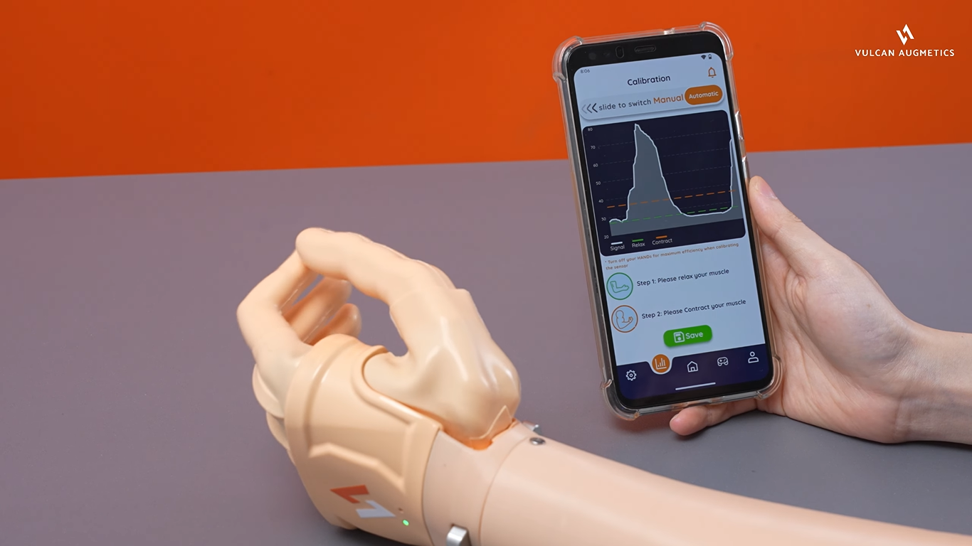
Accurate muscle signal assessment sits at the heart of upper-limb prosthetic care. From the very first myoelectric evaluation through to electrode placement, socket fitting, calibration, and long-term rehabilitation follow-up, clinicians depend on signal quality to make informed decisions at every stage. Yet in many clinicians today, that assessment still relies heavily on observation, experience, and subjective interpretation with limited access to measurable, real-time data. The Challenge: Limited Signal Visibility in Clinical Settings Surface EMG signals are inherently small and variable. They shift with limb position, tissue characteristics, socket fit, and muscle fatigue. When clinicians lack reliable, visible signal data, the consequences ripple across the entire care pathway: These gaps extend fitting timelines, reduce patient confidence, and can limit long-term prosthetic adoption. Real-Time Signal Visualization with the Vulcan Myoband The Vulcan Myoband addresses this directly by streaming real-time EMG and inertial motion (IMU) data through the Vulcan mobile app by putting objective signal information in the hands of both clinicians and patients during every stage of care. Worn as a sensor band around the residual limb, the Myoband captures muscle activation patterns, contraction timing, and arm movement dynamics simultaneously. Proprietary signal-processing algorithms convert raw biosignals into clear visual feedback, and the system automatically calculates and establishes activation thresholds calibrated to each individual’s muscle strength. The result is a dual-purpose interface designed for both clinical depth and patient clarity: This visual feedback loop supports a well-established principle in motor rehabilitation: when patients can see their muscle activity in real time, the brain reconnects with and learns to control those muscles more effectively. Clinical Applications and Benefits Real-time EMG and IMU visualization through the Vulcan app supports more informed, efficient prosthetic care across five key areas: 1. Objective myoelectric assessment Before a socket is even fabricated, clinicians can use the Myoband to verify whether a patient can generate stable, consistent muscle signals. This supports earlier and more confident prosthetic prescription decisions — reducing the risk of misclassifying candidates as unsuitable for myoelectric control. 2. Streamlined clinical workflow Signal capture, threshold visualization, and contraction analysis are all built into a single app. Clinics can conduct muscle assessments without investing in separate EMG diagnostic tools, reducing setup time and improving overall appointment efficiency. 3. Evidence-based calibration Visual feedback on contraction strength and response speed allows precise adjustment of threshold levels and control sensitivity. Clinicians can identify and minimize excessive muscle exertion, a common contributor to fatigue and long-term abandonment, with measurable data rather than subjective judgment. 4. Data-driven rehabilitation Stored signal and motion metrics can be reviewed over time, allowing therapists to track muscle activation trends, monitor recovery milestones, and adjust training plans based on objective progress data rather than recall alone. 5.Outcome measurement and reporting Quantitative biosignal data provides structured, reproducible metrics that contribute to clinical reporting and formal outcome assessments — supporting both individual patient care and broader service evaluation. From Experience-Based to Evidence-Based Prosthetic Fitting By making biosignal information visible, measurable, and shareable, the Vulcan ecosystem helps shift prosthetic fitting from a largely experience-dependent process toward a more data-guided clinical workflow. Real-time EMG and motion visualization gives patients a clearer understanding of their own control strategies, accelerates training, and builds the kind of long-term confidence that drives consistent prosthetic use. Learn more about Vulcan →

Real-Time EMG and IMU Visualization in Prosthetic Care: How the Vulcan App Supports Clinical Decision-Making Read More »