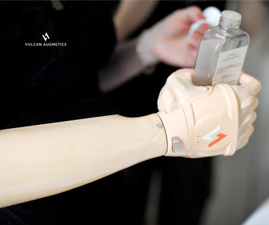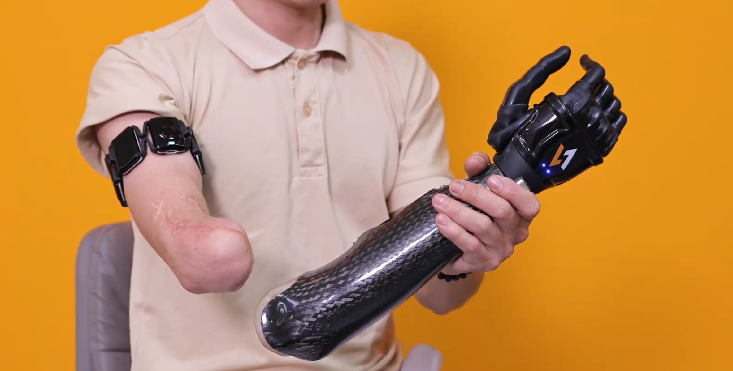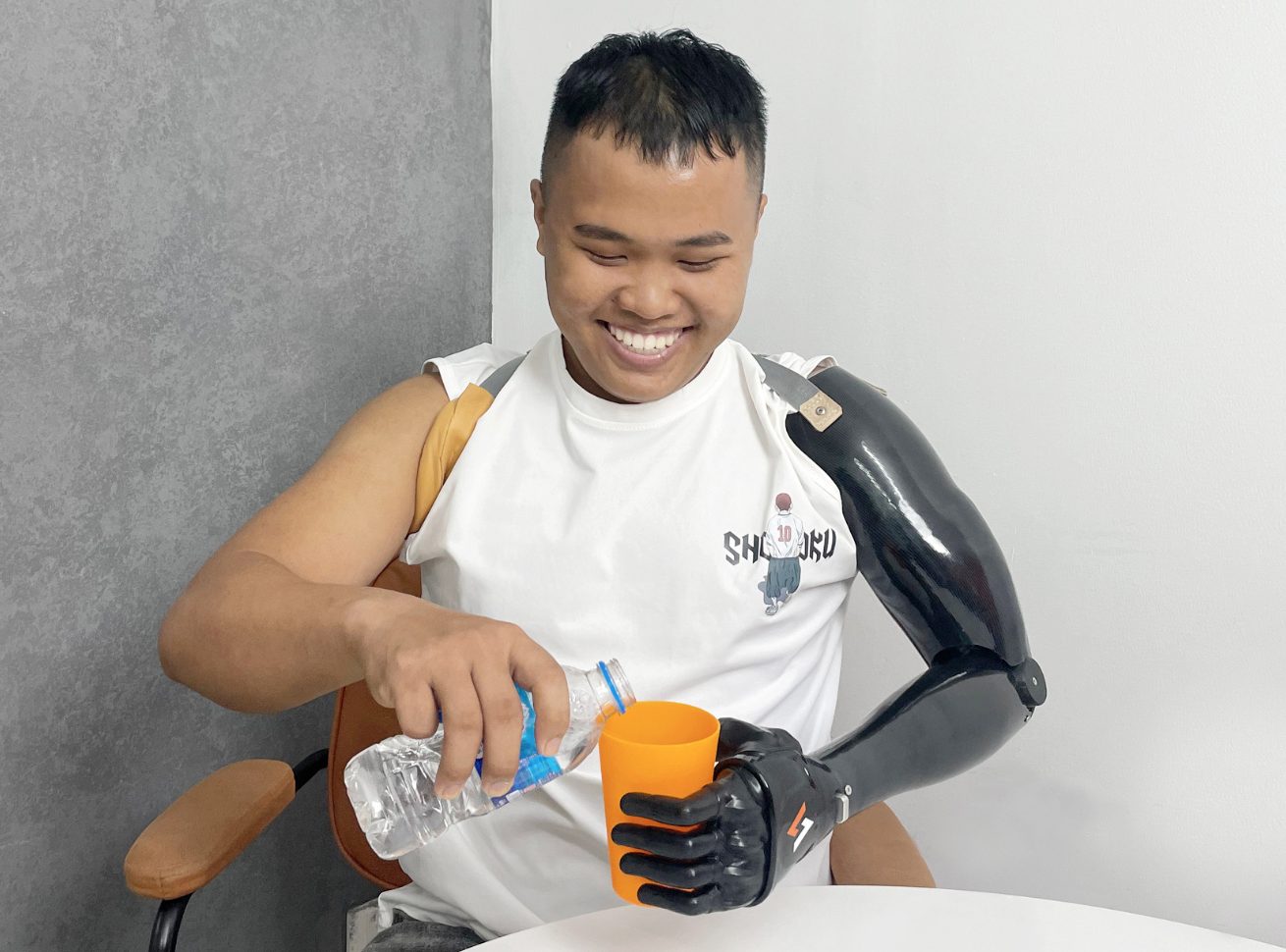

One of the less-discussed challenges in upperlimb prosthetic care is what happens to muscle tissue over time and what that means for myoelectric control.
For many patients, the residual limb doesn’t stay the same after amputation. Muscles that aren’t regularly activated begin to atrophy. Fatty and fibrous tissue accumulates. The signals that a prosthetic system needs to read become quieter, less stable, and harder to interpret. In some long-term cases or congenital presentations, those signals may be extremely faint from the outset.
Traditional electrode systems weren’t designed with this in mind and for a significant portion of the prosthetic population, that’s a real limitation.
What Happens to EMG Signals as Muscles Atrophy
Following amputation, the residual limb goes through a prolonged process of biological change. The most significant shifts typically occur in the first 6 to 18 months, but remodeling can continue indefinitely particularly in patients who don’t use a prosthesis early or who have limited physical activity.
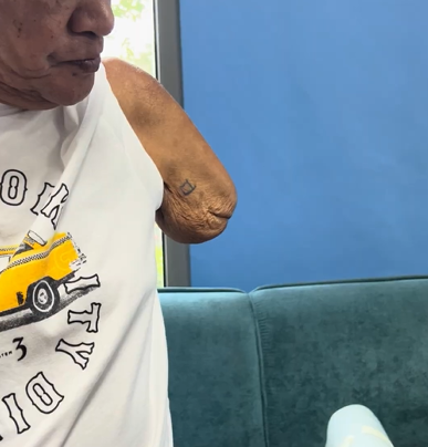
These changes affect EMG signal quality in several ways:
Signals become less stable
Atrophied muscle has fewer active motor units firing in a coordinated way. The result is a signal that fluctuates, making it difficult for patients to maintain a consistent contraction and for the system to interpret intent accurately.
Tissue changes increase impedance
As fatty and fibrous tissue accumulates between the muscle and the electrode, signal conduction attenuates. Electrode contact becomes less consistent, particularly as limb shape continues to change.
Noise becomes a bigger problem
When signal amplitude is low, the ratio of useful signal to background noise deteriorates. Crosstalk from adjacent muscle groups increases, and the risk of the system misreading a signal or missing one entirely goes up.
Why Conventional Electrode Systems Fall Short
Traditional dual-electrode setups depend on two things: accurate placement over a defined motor point, and signals strong enough to reliably cross a fixed threshold. In a healthy, recently-fitted patient, that’s a reasonable assumption.
In a patient with significant atrophy, neither condition may hold. Motor points may no longer be clearly defined. Signals may never consistently reach the threshold needed to trigger a command. The system that worked at fitting may become unreliable months later as the limb continues to change.
For long-term amputees, non-users returning to prosthetic care, or individuals with congenital limb differences, this creates a meaningful barrier to myoelectric control — not because the patient can’t generate signals, but because the system can’t detect and interpret the ones they have.
How Vulcan Approaches It Differently
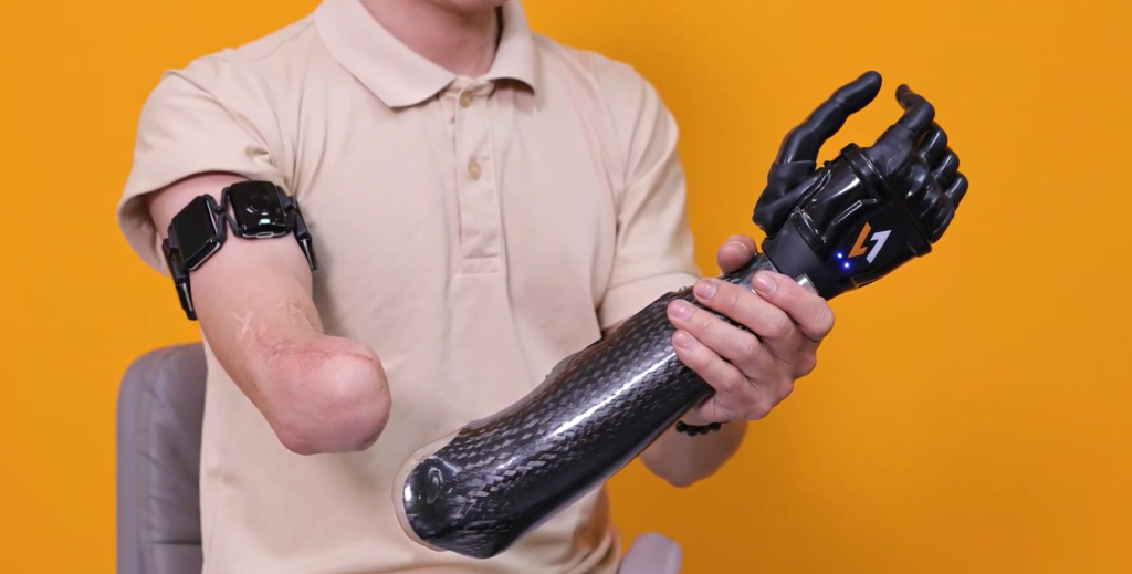
Rather than requiring a strong signal from a fixed location, the Vulcan Myoband is designed to work with what’s available across the full residual limb.
The Myoband encircles the limb with multiple sensors, so it isn’t dependent on a single site holding up over time. If muscle activity in one area is reduced, the system continues to draw on signal from elsewhere around the limb, meaning a single atrophied or fibrotic region doesn’t determine whether control is possible.
Reading Signal, Not Just Strength
Conventional systems work on a simple rule: if the signal crosses a set strength, trigger a command. For patients with weak or atrophied muscle signals, that level may never be consistently reachable.
The Vulcan Myoband is built to recognize meaningful muscle activity even when it’s faint, calibrating to what each patient’s signal actually looks like, rather than expecting every patient to produce the same signal strength.
Built for Faint and Inconsistent Signals
Patients with significant atrophy often generate muscle activity too subtle for conventional electrodes to register. The Myoband is designed to pick up on activity at this level, even when individual signals are small or inconsistent.
This gives clinicians a clearer picture of where viable muscle activity exists across the residual limb, useful information when deciding which sites to focus on during training, especially in complex or long-term cases.
A Three-Step Clinical Protocol for Atrophy Cases
For patients with muscle weakness or long-term limb loss, Vulcan’s approach follows a structured sequence:
1. Calibration
The Vulcan app learns the patient’s current contraction levels during initial setup, even if those contractions are extremely faint. Rather than requiring the patient to meet a fixed standard, the system is calibrated individually, starting from where the patient’s signal actually is.
2. Visual feedback
The patient can see their muscle activity in on screen. This isn’t just reassuring, it’s clinically meaningful. Visual feedback helps the brain reconnect with and learn to control muscles that may have had limited use for months or years, supporting the neuromuscular re-engagement that underpins effective prosthetic training.
3. Physical conditioning
Alongside device training, daily massage and isometric exercises help maintain remaining muscle fiber density and improve local blood flow. Preserving what muscle tissue remains makes a measurable difference to long-term signal quality and control consistency.
Why This Matters for Clinical Practice
Muscle atrophy is not a niche presentation. It affects long-term amputees, late fitters, patients who’ve had previous prosthetic abandonment, and individuals with congenital limb differences. It’s also a progressive condition, meaning patients who are well-controlled at fitting may become harder to manage over time if the system can’t adapt.
A control approach that reads signal patterns rather than raw amplitude, captures data across the full limb rather than fixed points, and adapts its baseline to the individual patient is better positioned to serve this population both at the point of fitting and across the years that follow.