
Before a patient can successfully control a myoelectric prosthesis, clinicians must first solve one of the most critical and often unpredictable aspects of the fitting process: acquiring reliable EMG signals.
While prosthetic technology continues to evolve, the quality of myoelectric control still depends heavily on what happens during signal acquisition. The effectiveness of control training, calibration, and long-term device performance all begin with the ability to consistently identify and capture usable muscle activity.
Why Is EMG Signal Acquisition Challenging?
In theory, acquiring a usable EMG signal is straightforward: place electrodes over the target muscle, ask the patient to contract, observe the output. In practice, the process is confounded by a range of patient-specific, physiological, and anatomical variables that make no two fittings alike.
1. Residual Limb Variability
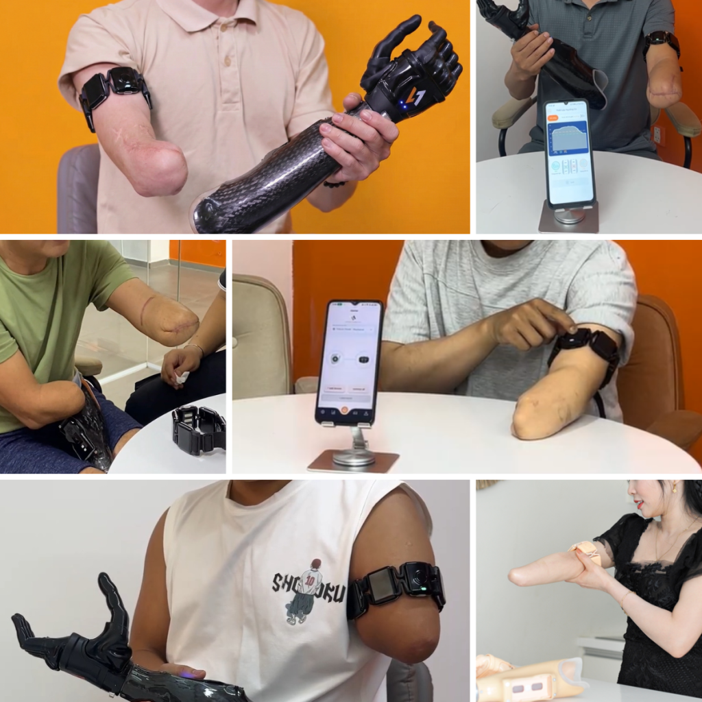
Limb length, tissue composition, skin condition, and scar tissue distribution differ significantly between patients and within the same patient over time as the limb matures post-amputation.
2. Muscle Atrophy and Reduced Signal Strength
Patients with long-term limb loss often present with significantly reduced muscle mass in the residual limb. Atrophied muscle produces weaker signals with lower signal-to-noise ratios, making reliable acquisition technically demanding.
3. Scar Tissue Interference
Post-surgical scar tissue increases impedance at the skin surface, attenuating signal amplitude and introducing inconsistency in electrode contact quality, particularly problematic for traditional socket-embedded electrodes.
4. Signal Crosstalk
In short residual limbs or limbs with limited tissue differentiation, electrical activity from adjacent muscles can contaminate the target signal, producing false activations and unpredictable control responses.
5. Variability in Voluntary Control
New prosthetic users often lack consistent voluntary muscle activation patterns. Signals vary in amplitude, timing, and repeatability between sessions, particularly in the early weeks post-fitting.
6. Dynamic Changes During Daily Use
Traditional socket-embedded electrodes are positioned for a specific limb configuration. Limb volume changes from perspiration, weight fluctuation, or tissue adaptation can shift electrode-skin contact and degrade signal quality between fittings.
These variables do not present in isolation. A typical complex patient may exhibit weak baseline signals, compromised tissue quality, and limited voluntary control simultaneously, requiring the CPO to manage all three in a single fitting session.
How Poor EMG Signal Acquisition Affects Clinical Outcomes
Signal acquisition problems are not always obvious during fitting appointments. Patients may demonstrate acceptable control while seated in a controlled clinical environment yet struggle significantly once they return to the unpredictable demands of daily life.
Poor signal quality can contribute to:
- Increased recalibration frequency
- Longer training periods
- Reduced control reliability
- Greater clinician followup burden
- Decreased patient confidence
- Reduced daily wear time
- Higher risk of prosthesis abandonment
Every unsuccessful fitting carries hidden costs, not only in clinician time and resources but also in the patient’s trust in the technology itself.
A Systematic Approach to EMG Signal Assessment
Managing signal acquisition complexity begins before the electrode is placed. A structured pre-fitting assessment protocol reduces the likelihood of downstream control problems and gives the CPO a reproducible framework for evaluating each patient’s signal landscape.
1. Residual Limb Mapping
Before electrode placement, systematically assess the residual limb for tissue quality, scar tissue location, muscle belly palpation, and approximate signal site candidates. Document findings for reference across sessions. This step is frequently abbreviated under time pressure — at significant downstream cost.
2. Baseline Signal Characterization
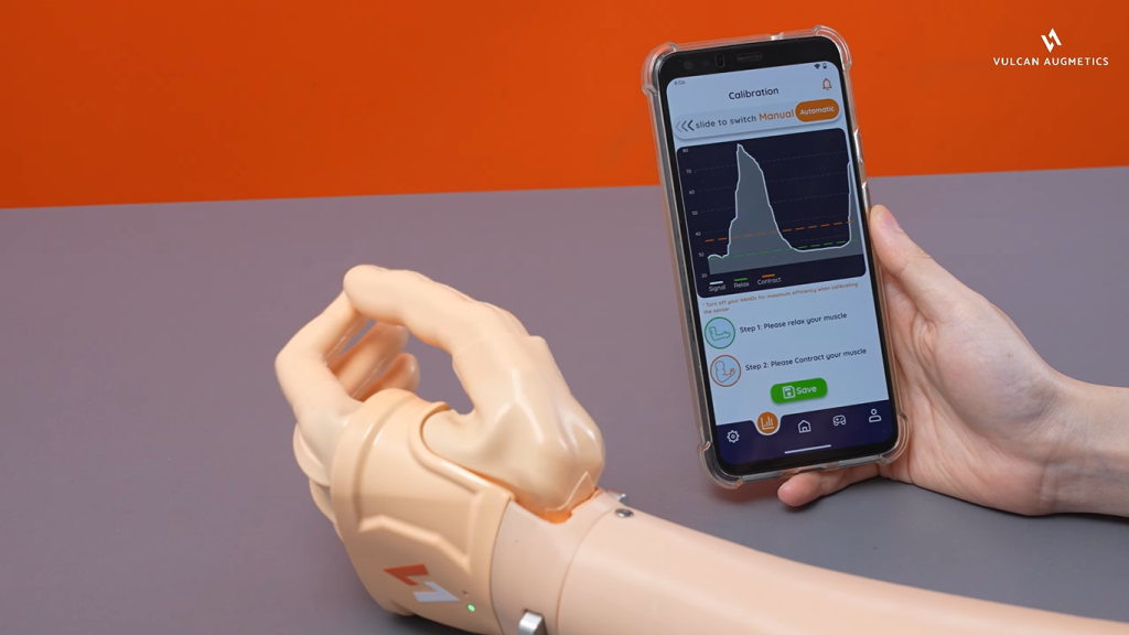
Use real-time EMG visualization to establish baseline signal amplitude, resting noise levels, and voluntary contraction quality across candidate sites before committing to electrode placement. Compare multiple sites systematically rather than relying on the first adequate signal identified.
3. Dynamic Signal Testing
Assess signal quality under movement conditions, not just at rest. Have the patient perform arm movements, elevation changes, and load-bearing tasks while monitoring signal consistency. A signal that is stable in static testing often degrades significantly under dynamic conditions.
4. Patient-Specific Threshold Calibration
Calibrate activation thresholds to the individual patient’s signal profile — not to a population average. Patients with weaker signals require lower thresholds and higher gain settings; patients with strong signals or crosstalk may need more restrictive thresholds to prevent false activations.
5. Session-to-Session Reassessment
At the second fitting session, re-evaluate signal quality with the same protocol used in session one. Significant variation between sessions is diagnostically important, it identifies patients whose signal profile is not yet stable and who will require ongoing monitoring and recalibration.
How Multi-Sensor Coverage Changes the Acquisition Equation
The choice of control approach depends on the patient’s functional goals and signal profile. But regardless of which approach a clinician selects, the quality of EMG signal acquisition upstream determines how well any system will perform. Advanced control technologies depend on consistent and repeatable signal input to function as intended. When the underlying signal is weak, inconsistent, or contaminated by crosstalk, no control layer can fully compensate.
Traditional myoelectric systems rely on precise placement of one or two electrodes over specific muscle bellies. The clinician must palpate the residual limb while the patient performs repeated contractions to locate a single viable motor point: a process clinicians often describe as “myosite hunting.” In ideal anatomy, this works well. In residual limbs with scar tissue, atrophy, or limited muscle differentiation, it can consume a disproportionate share of fitting time before device viability is even confirmed.
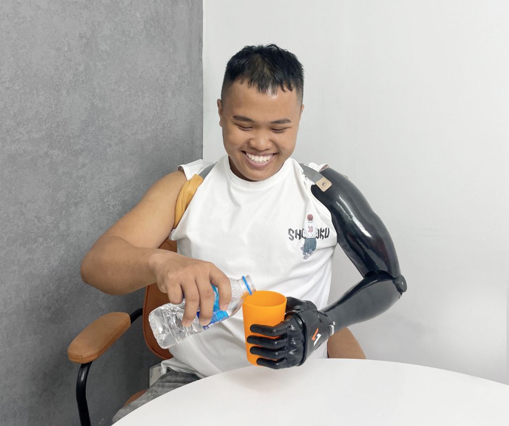
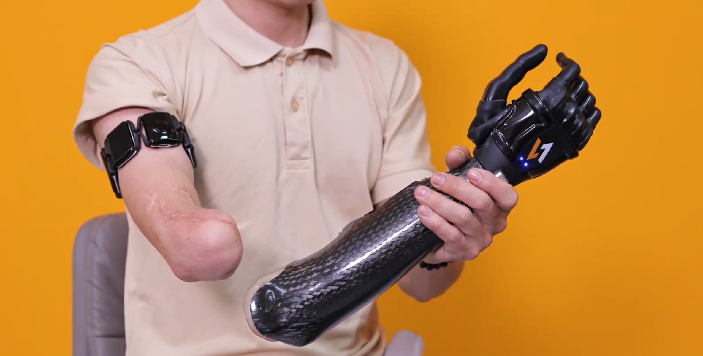
The Vulcan control system calibrates automatically to each patient’s individual muscle activity through the Vulcan app in roughly 60 seconds. Commands are transmitted wirelessly to the prosthetic hand, with no embedded wiring inside the socket to maintain.
What Consistent Signal Acquisition Makes Possible
The clinical benefits of reliable EMG signal acquisition extend well beyond fitting day. When signal quality is stable and reproducible, everything downstream becomes more manageable: control training is faster, patient confidence builds earlier, recalibration frequency drops, and long-term device adoption improves.
The goal of signal acquisition is not to obtain the strongest signal. It is to obtain the most reliable signal for that patient, one that will perform consistently across the full range of conditions their daily life presents. That requires a systematic approach, the right assessment tools, and a sensing architecture designed for real-world variability rather than clinical ideal conditions.
See How Vulcan Approaches Signal Acquisition
Explore how the Myoband’s multi-sensor architecture simplifies clinical fitting across complex patient profiles.
Fitting sessions test signal quality under static, controlled conditions. Daily use introduces variables: perspiration, limb volume changes, socket movement, fatigue that can shift electrode contact or degrade signal amplitude. A signal that looks stable during a seated clinic assessment may not hold up during a full day of real-world activity.