CPO Resources

A close-up side profile of an elderly individual with an upper-limb amputation above the elbow. The residual limb shows visible skin wrinkling and signs of muscle atrophy. A small, simple square tattoo is visible on the side of the limb.

When Muscles Change: How Vulcan Supports Patients With Muscle Atrophy and Long-Term Limb Loss

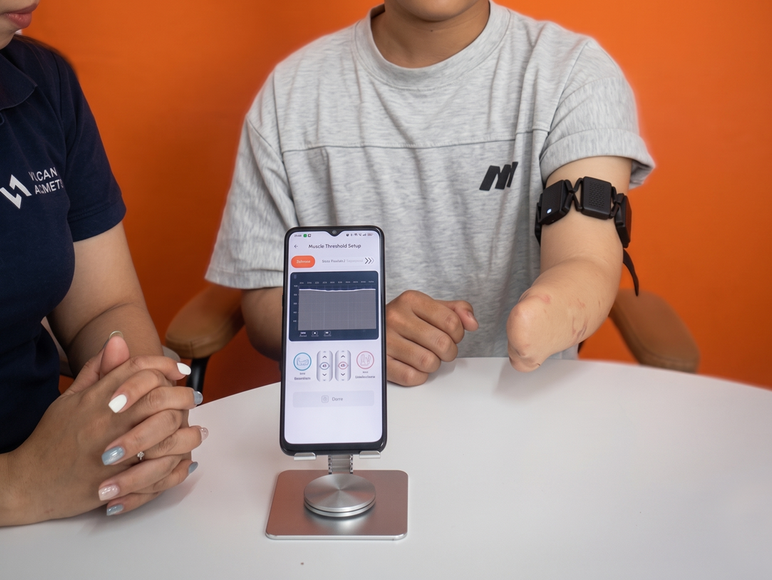
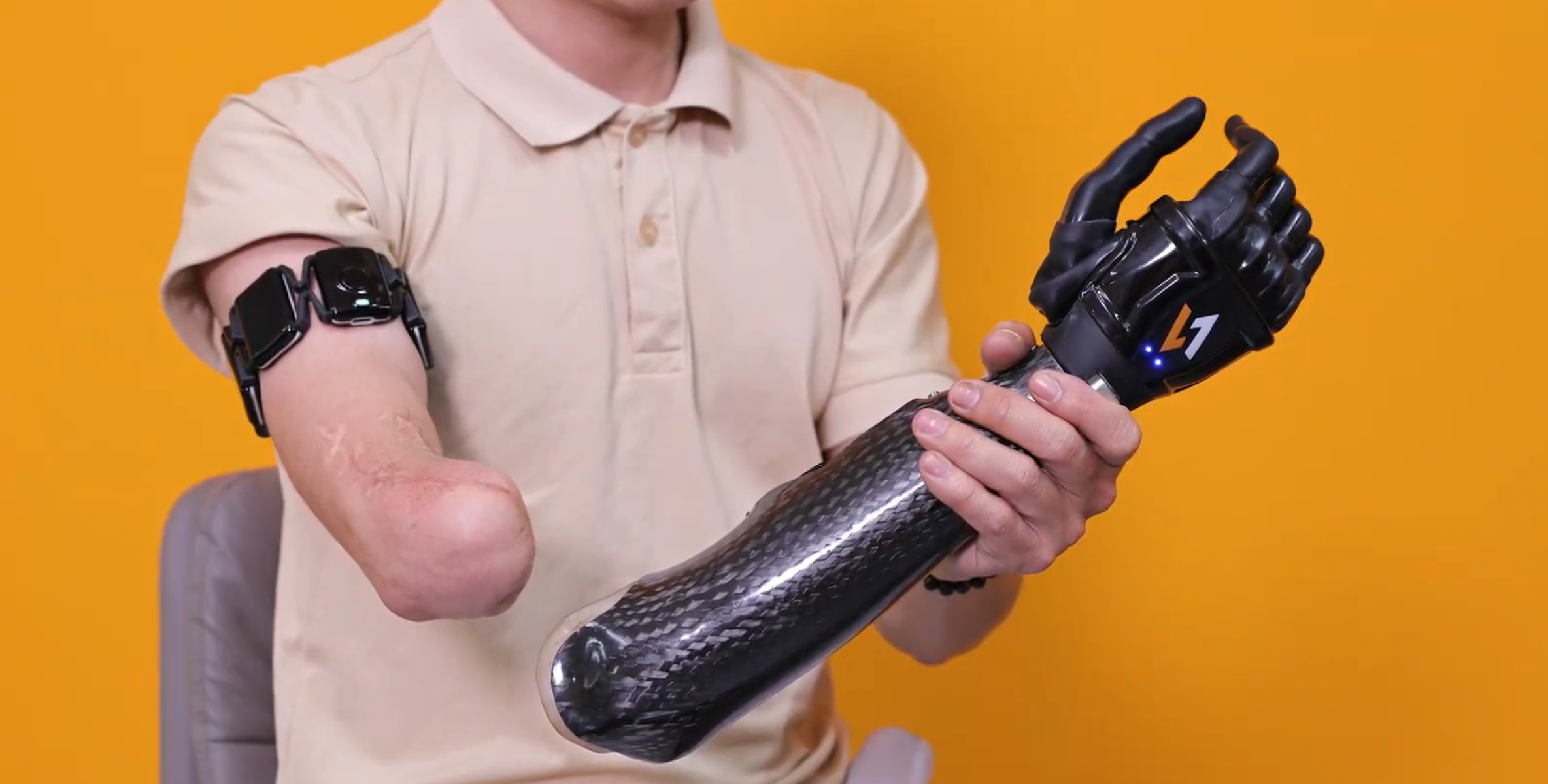
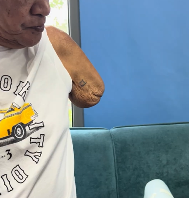
One of the less-discussed challenges in upperlimb prosthetic care is what happens to muscle tissue over time and what that means for myoelectric control. For many patients, the residual limb doesn’t stay the same after amputation. Muscles that aren’t regularly activated begin to atrophy. Fatty and fibrous tissue accumulates. The signals that a prosthetic system needs to read become quieter, less stable, and harder to interpret. In some long-term cases or congenital presentations, those signals may be extremely faint from the outset. Traditional electrode systems weren’t designed with this in mind and for a significant portion of the prosthetic population, that’s a real limitation. What Happens to EMG Signals as Muscles Atrophy Following amputation, the residual limb goes through a prolonged process of biological change. The most significant shifts typically occur in the first 6 to 18 months, but remodeling can continue indefinitely particularly in patients who don’t use a prosthesis early or who have limited physical activity. These changes affect EMG signal quality in several ways: Signals become less stable Atrophied muscle has fewer active motor units firing in a coordinated way. The result is a signal that fluctuates, making it difficult for patients to maintain a consistent contraction and for the system to interpret intent accurately Tissue changes increase impedance As fatty and fibrous tissue accumulates between the muscle and the electrode, signal conduction attenuates. Electrode contact becomes less consistent, particularly as limb shape continues to change. Noise becomes a bigger problem When signal amplitude is low, the ratio of useful signal to background noise deteriorates. Crosstalk from adjacent muscle groups increases, and the risk of the system misreading a signal or missing one entirely goes up. Why Conventional Electrode Systems Fall Short Traditional dual-electrode setups depend on two things: accurate placement over a defined motor point, and signals strong enough to reliably cross a fixed threshold. In a healthy, recently-fitted patient, that’s a reasonable assumption. In a patient with significant atrophy, neither condition may hold. Motor points may no longer be clearly defined. Signals may never consistently reach the threshold needed to trigger a command. The system that worked at fitting may become unreliable months later as the limb continues to change. For long-term amputees, non-users returning to prosthetic care, or individuals with congenital limb differences, this creates a meaningful barrier to myoelectric control — not because the patient can’t generate signals, but because the system can’t detect and interpret the ones they have. How Vulcan Approaches It Differently Rather than requiring a strong signal from a fixed location, the Vulcan Myoband is designed to work with what’s available and to find it across the full residual limb. Vulcan Myoband encircles the limb with multiple sensors. If one area is atrophied or fibrotic, the system scans across all sensor sites and prioritizes data from wherever the most active muscle tissue remains. This spatial flexibility means the system isn’t dependent on a single motor point holding up over time. Pattern recognition over amplitude thresholds Conventional systems operate on a simple rule: if the signal exceeds a set value, trigger a command. For patients with weak signals, that threshold may never be consistently reachable. The Vulcan system learns the shape of each patient’s signal rather than just its amplitude. This shifts the control logic from “how strong is the signal” to “what does this signal mean for this patient.” Micro-activation detection and spatial mapping Advanced noise filtering allows the system to detect very small muscle activations that would be invisible to conventional electrodes. Spatial mapping builds a distribution picture of activity across the residual limb, helping clinicians identify the most viable muscle sites to target during training, even in complex or long-term cases. A Three-Step Clinical Protocol for Atrophy Cases For patients with muscle weakness or long-term limb loss, Vulcan’s approach follows a structured sequence: 1. Calibration The Vulcan app learns the patient’s current contraction levels during initial setup — even if those contractions are extremely faint. Rather than requiring the patient to meet a fixed standard, the system adapts its baseline to where the patient actually is. 2. Visual feedback The patient can see their muscle activity in real time on screen. This isn’t just reassuring, it’s clinically meaningful. Visual feedback helps the brain reconnect with and learn to control muscles that may have had limited use for months or years, supporting the neuromuscular re-engagement that underpins effective prosthetic training. 3. Physical conditioning Alongside device training, daily massage and isometric exercises help maintain remaining muscle fiber density and improve local blood flow. Preserving what muscle tissue remains makes a measurable difference to long-term signal quality and control consistency. Why This Matters for Clinical Practice Muscle atrophy is not a niche presentation. It affects long-term amputees, late fitters, patients who’ve had previous prosthetic abandonment, and individuals with congenital limb differences. It’s also a progressive condition, meaning patients who are well-controlled at fitting may become harder to manage over time if the system can’t adapt. A control approach that reads signal patterns rather than raw amplitude, captures data across the full limb rather than fixed points, and adapts its baseline to the individual patient is better positioned to serve this population both at the point of fitting and across the years that follow.

When Muscles Change: How Vulcan Supports Patients With Muscle Atrophy and Long-Term Limb Loss Read More »

A clinician and a patient with a transradial amputation evaluating real-time EMG signals. The patient wears a Vulcan wireless myoband on the residual limb, while a smartphone on a stand displays a visual graph of muscle activation thresholds.

Real-Time EMG and IMU Visualization in Prosthetic Care: How the Vulcan App Supports Clinical Decision-Making

Accurate muscle signal assessment sits at the heart of upper-limb prosthetic care. From the very first myoelectric evaluation through to electrode placement, socket fitting, calibration, and long-term rehabilitation follow-up, clinicians depend on signal quality to make informed decisions at every stage. Yet in many clinicians today, that assessment still relies heavily on observation, experience, and subjective interpretation with limited access to measurable, real-time data. The Challenge: Limited Signal Visibility in Clinical Settings Surface EMG signals are inherently small and variable. They shift with limb position, tissue characteristics, socket fit, and muscle fatigue. When clinicians lack reliable, visible signal data, the consequences ripple across the entire care pathway: These gaps extend fitting timelines, reduce patient confidence, and can limit long-term prosthetic adoption. Real-Time Signal Visualization with the Vulcan Myoband The Vulcan Myoband addresses this directly by streaming real-time EMG and inertial motion (IMU) data through the Vulcan mobile app by putting objective signal information in the hands of both clinicians and patients during every stage of care. Worn as a sensor band around the residual limb, the Myoband captures muscle activation patterns, contraction timing, and arm movement dynamics simultaneously. Proprietary signal-processing algorithms convert raw biosignals into clear visual feedback, and the system automatically calculates and establishes activation thresholds calibrated to each individual’s muscle strength. The result is a dual-purpose interface designed for both clinical depth and patient clarity: This visual feedback loop supports a well-established principle in motor rehabilitation: when patients can see their muscle activity in real time, the brain reconnects with and learns to control those muscles more effectively. Clinical Applications and Benefits Real-time EMG and IMU visualization through the Vulcan app supports more informed, efficient prosthetic care across five key areas: 1. Objective myoelectric assessment Before a socket is even fabricated, clinicians can use the Myoband to verify whether a patient can generate stable, consistent muscle signals. This supports earlier and more confident prosthetic prescription decisions — reducing the risk of misclassifying candidates as unsuitable for myoelectric control. 2. Streamlined clinical workflow Signal capture, threshold visualization, and contraction analysis are all built into a single app. Clinics can conduct muscle assessments without investing in separate EMG diagnostic tools, reducing setup time and improving overall appointment efficiency. 3. Evidence-based calibration Visual feedback on contraction strength and response speed allows precise adjustment of threshold levels and control sensitivity. Clinicians can identify and minimize excessive muscle exertion, a common contributor to fatigue and long-term abandonment, with measurable data rather than subjective judgment. 4. Data-driven rehabilitation Stored signal and motion metrics can be reviewed over time, allowing therapists to track muscle activation trends, monitor recovery milestones, and adjust training plans based on objective progress data rather than recall alone. 5.Outcome measurement and reporting Quantitative biosignal data provides structured, reproducible metrics that contribute to clinical reporting and formal outcome assessments — supporting both individual patient care and broader service evaluation. From Experience-Based to Evidence-Based Prosthetic Fitting By making biosignal information visible, measurable, and shareable, the Vulcan ecosystem helps shift prosthetic fitting from a largely experience-dependent process toward a more data-guided clinical workflow. Real-time EMG and motion visualization gives patients a clearer understanding of their own control strategies, accelerates training, and builds the kind of long-term confidence that drives consistent prosthetic use. Learn more about Vulcan →

Real-Time EMG and IMU Visualization in Prosthetic Care: How the Vulcan App Supports Clinical Decision-Making Read More »

An arm amputee man wearing two black Vulcan prosthetic hands on his both stumps hold his wife's hands in an orange room

Optimizing the Myoelectric Clinical Workflow: Benefits of the Vulcan Myoband

The Vulcan Myoband is designed not only to improve prosthetic control for patients, but also to simplify the clinical workflow for healthcare providers and partners across the prosthetic ecosystem. By combining wearable biosignal sensing, adaptive signal processing, and wireless control architecture, the system delivers practical advantages for patients, clinicians, physicians, and distributors. Improved Control for Patients Instead of requiring strong or perfectly isolated contractions, the system recognizes subtle changes in muscle activation. This allows more patients to achieve functional control with less effort and a shorter learning curve. Simplified Clinical Fitting Workflow For prosthetists and clinical technicians, the Myoband’s flexibility can help reduce setup time, simplify adjustments, and improve fitting efficiency in daily clinical practice by: Expanded Opportunities for Rehabilitation Teams 1. Rapid, Patient-Specific Calibration The Vulcan ecosystem can perform automatic signal calibration in under 60 seconds, identifying individualized muscle contraction and relaxation thresholds. Clinicians can then fine-tune parameters through the mobile app to optimize control sensitivity and responsiveness based on each patient’s rehabilitation progress. This technology helps establish a personalized control profile quickly, reducing the time required to initiate functional training. 2. Faster Motor Learning and Functional Engagement Because control is based on natural muscle activation threshold recognition, many patients can begin basic task-oriented training soon after fitting. In clinical practice, some patients are able to perform simple functional exercises, such as holding a cup, writing, grasping paper, or peeling fruit, after a short 30-minute-familiarization session. Earlier functional engagement can support motor relearning, confidence building, and motivation during rehabilitation. 3. Potential for Earlier Prosthetic Adoption By simplifying signal detection and accelerating control training, the Myoband may help rehabilitation teams introduce myoelectric prostheses earlier in the recovery timeline, when appropriate from a medical standpoint. Earlier exposure to functional prosthetic use can contribute to: Scalable Solutions for Distributors and Providers 1. Easy Integration Into Existing Prosthetic Workflows The Myoband’s wearable, modular architecture and wireless connectivity allow seamless integration with current prosthetic systems, reducing technical barriers during setup and configuration. 2. Scalable Solution for Diverse Markets A streamlined fitting process and external sensor design help lower operational complexity, enabling clinics to deliver myoelectric solutions more efficiently while supporting a wider range of patient needs. The Vulcan ecosystem also supports consistent service delivery across some different regions, practice settings, and patient populations, as a result, providers can deliver reliable myoelectric solutions with greater efficiency and scalability. Explore Clinical Integration To review technical specifications, request system documentation, or discuss integrating the Vulcan ecosystem into your clinical practice, please contact our partnership team. [Contact Vulcan Provider Support]

Optimizing the Myoelectric Clinical Workflow: Benefits of the Vulcan Myoband Read More »

Who Can Use a Myoelectric Hand? Patient Suitability and the Vulcan Solution

A primary consideration in upper-limb rehabilitation is determining a patient’s suitability for a myoelectric prosthesis. Traditionally, successful myoelectric control requires a residual limb with specific characteristics, particularly strong and isolated electromyographic (EMG) signals. The Vulcan Myoband provides an alternative approach to myoelectric control. By utilizing a flexible, wireless Vulcan Myoband instead of standard rigid socket electrodes, the system is designed to accommodate a broader range of anatomical conditions and signal presentations. The Vulcan system is applicable for multiple upper-limb amputation levels. It is currently indicated for patients undergoing rehabilitation for: Clinical Note: The Vulcan Myoband and prosthetic hand system are not currently indicated for partial hand or individual finger amputations. Residual Limb and EMG Signal Considerations  In patients with complex residual limb conditions, identifying stable signal locations can be difficult and may require repeated calibration. Myoband addresses this challenge through high-sensitivity EMG sensors distributed around the forearm, as well as a smart algorithm developed by Vulcan, allowing the system to capture muscle activity from several channels simultaneously and reliably. This approach can improve signal detection reliability in patients with less predictable signal patterns, allowing it to adapt to various anatomical presentations where traditional single-site electrodes often fail. 1. Scar Tissue and Skin Grafts Scar tissue typically exhibits high electrical resistance, which can attenuate EMG signals and impede detection by standard single-point sensors. The Vulcan Control Solution: If localized scar tissue causes signal attenuation at one site, the algorithm relies on data acquired from adjacent sensors to capture the overall muscle activation pattern. 2. Bone Overgrowth and Irregular Contours Rigid electrodes require flat, continuous contact with the skin. Irregular stump shapes or bony protrusions can cause pressure points, discomfort, and inconsistent sensor contact (motion artifacts). The Vulcan Control Solution: Vulcan Myoband uses a flexible, modular elastic structure. This design allows the sensors to conform to the limb’s natural contours, maintaining contact without applying localized pressure on prominent bones. 3. Bilateral Amputations Clinical Case 3 – Bilateral Amputations [ Watch the video here] Extreme difficulty for the patient to don/doff devices and perform daily calibrations independently. The Vulcan Solution: The band design allows patients to slide the device on using their other stump. Software profiles are saved per limb, making synchronized bilateral control intuitive. 4. Osseointegration  Requires a system that doesn’t interfere with the percutaneous implant site while maintaining high-fidelity control. The Vulcan Solution: Since the bone is fixed, muscle movement is more predictable. Myoband captures these stable muscle contractions non-invasively, providing a high-speed interface that matches the direct-to-bone stability. For clinicians and orthotists, utilizing Vulcan Myoband approach simplifies the socket fabrication process by removing embedded wiring. Functionally, it provides a viable control option for patients whose signal patterns or anatomical features previously excluded them from using standard myoelectric systems. For more information regarding system specifications, user manuals, or to view additional case studies demonstrating the Vulcan Myoband in various clinical scenarios, please visit our resource center. Click here

Who Can Use a Myoelectric Hand? Patient Suitability and the Vulcan Solution Read More »

A person with a below-the-elbow amputation wearing the Vulcan Myoband—a black, segmented multi-sensor armband—on their upper arm. They are holding a sleek, carbon-fiber prosthetic hand (marked with the 'V1' logo) to demonstrate the connection between the sensors and the device against a bright yellow background.

Optimizing Myoelectric Hand Control: How the Vulcan Myoband Overcomes Clinical Fitting Challenges

After an upper-limb amputation, receiving a myoelectric prosthetic hand can take one to six months, even after insurance approval or out-of-pocket agreement. The fitting process alone typically requires three to four clinic visits, depending on the complexity of the limb condition and the fabrication workflow. Yet despite this time and effort, myoelectric fittings still fail in many clinics. So why does fitting still take so long? Several factors commonly slow down the process: The EMG Signal Challenge Myoelectric prosthetic control relies on electromyographic (EMG) signals, which are generated when residual muscles contract. After an upperlimb amputation, the remaining muscles in the forearm or upper arm can still produce these signals when a patient attempts to move the missing limb. However, the characteristics of these signals vary widely from patient to patient. The biology of the residual limb and the original surgical procedure play a major role in determining how usable these signals are for prosthetic control. Several factors influence EMG signal quality, including: In addition, surgical reconstruction can alter the natural structure of the muscle system. Depending on the procedure, residual muscles may be reattached to bone or tendon, which can change how neuromuscular signals are generated and transmitted. As a result, EMG signals in an amputated limb often differ from those in an intact limb. They typically show: Surface EMG sensors must detect these signals through several biological layers, including muscle tissue, connective tissue, subcutaneous fat, and skin, which further attenuate and filter the electrical activity. Because of these physiological factors, sensor placement becomes critical. Clinicians must identify the most active muscle regions and optimize electrode placement based on signal amplitude, soft tissue thickness, and stability within the prosthetic socket. Why Electrode Placement Is So Difficult Most traditional myoelectric systems requiring electrodes to be placed on two opposing muscle groups. For reliable detection, electrodes must be positioned precisely on the muscle belly and aligned with the direction of the muscle fibers. During fitting, prosthetists often ask patients to perform repeated muscle contractions while adjusting electrode locations to find signals that are both strong and distinguishable. In practice, this process can involve extensive repositioning and testing, sometimes referred to clinically as “myosite hunting.” Even when optimal placement is achieved in the clinic, maintaining stable signals in daily life remains challenging. Factors such as electrode displacement, socket pressure changes, sweat, and natural biological changes in the residual limb can all affect signal quality. Socket Limitations In conventional prosthetic systems, electrodes are typically embedded inside the socket, which creates additional constraints. Socket design must balance structural strength, comfort, and space for electronic components. At the same time, electrode locations become fixed once the socket is fabricated. However, residual limb volume can fluctuate throughout the day, and small shifts in socket position during daily movement can alter electrode alignment. When signal quality changes, patients may require re-adjustments or even a new socket, extending the fitting timeline and sometimes leading to frustration or abandonment of the prosthesis. The Result: A Trial-and-Error Process Because EMG signals vary between individuals and depend heavily on precise electrode placement, the fitting process often becomes iterative and time-consuming. Multiple adjustments, test fittings, and recalibrations may be needed before achieving stable control. For patients eager to regain independence, these delays can be discouraging. For clinicians, they represent one of the most persistent challenges in modern myoelectric prosthetic care. How the Vulcan Myoband Addresses Common EMG Signal Detection Challenges Reliable EMG signal detection remains one of the most common challenges in myoelectric prosthetic control. In clinical practice, obtaining stable signals can be difficult, especially for patients with complex residual limb conditions. Supporting Patients With Weak or Complex EMG Signals The challenge: Many patients struggle to produce strong, clearly separated EMG signals due to factors such as: The Vulcan Solution: Instead of requiring strong or highly isolated contractions, the Myoband detects subtle increases in muscle activation above the resting state to establish basic open-close control of the prosthetic hand, based on a threshold recognition technology. As a result, patients may experience: Scar tissue, thick subcutaneous fat, or skin grafts, common in burn or trauma patients, can weaken EMG signals, making it difficult to identify two reliable muscle sites for traditional dual-electrode control on the residual limb. The Myoband overcomes this by being worn around the upper arm and using multiple sensors to capture overall muscle activation across a wider area. Even if one region produces weaker muscle signals due to scarring or reduced sensitivity, the system can still detect activity from surrounding healthy muscle tissue. Reduced Dependence on Precise Electrode Placement The Challenge: Traditional myoelectric systems often require precise electrode placement over specific muscle sites. Small positional changes can significantly affect signal quality. The Vulcan Solution: The Myoband reduces this dependency through multi-channel EMG sensing distributed around the upper arm, capturing signals from several muscle regions simultaneously. This design offers several practical advantages: Stability During Daily Movement The Challenge: During everyday activities, prosthetic sockets may shift slightly on the residual limb, a phenomenon known as pistoning. When electrodes are embedded inside the socket, this movement can disrupt signal detection. The Vulcan Solution: Because the Myoband is worn directly on the arm, the sensors maintain more stable contact with the skin.  The Myoband also integrates an IMU (Inertial Measurement Unit) that detects arm position and orientation. This helps distinguish between intentional muscle activation and postural contractions. For example: The system is also designed to handle environmental factors such as sweat and changes in skin impedance, using adaptive algorithms that maintain stable activation thresholds and reduce unintended hand movements.

Optimizing Myoelectric Hand Control: How the Vulcan Myoband Overcomes Clinical Fitting Challenges Read More »